Iodine »
PDB 8ssv-9ku6 »
8u0r »
Iodine in PDB 8u0r: The Crystal Structure of Protein A21, A Component of the Conserved Poxvirus Entry-Fusion Complex
Protein crystallography data
The structure of The Crystal Structure of Protein A21, A Component of the Conserved Poxvirus Entry-Fusion Complex, PDB code: 8u0r
was solved by
U.Diesterbeck,
A.G.Gittis,
D.N.Garboczi,
B.Moss,
with X-Ray Crystallography technique. A brief refinement statistics is given in the table below:
| Resolution Low / High (Å) | 39.31 / 2.30 |
| Space group | P 1 21 1 |
| Cell size a, b, c (Å), α, β, γ (°) | 46.86, 70.06, 78.95, 90, 95.29, 90 |
| R / Rfree (%) | 21.1 / 23.9 |
Other elements in 8u0r:
The structure of The Crystal Structure of Protein A21, A Component of the Conserved Poxvirus Entry-Fusion Complex also contains other interesting chemical elements:
| Sodium | (Na) | 2 atoms |
| Chlorine | (Cl) | 9 atoms |
Iodine Binding Sites:
Pages:
>>> Page 1 <<< Page 2, Binding sites: 11 - 12;Binding sites:
The binding sites of Iodine atom in the The Crystal Structure of Protein A21, A Component of the Conserved Poxvirus Entry-Fusion Complex (pdb code 8u0r). This binding sites where shown within 5.0 Angstroms radius around Iodine atom.In total 12 binding sites of Iodine where determined in the The Crystal Structure of Protein A21, A Component of the Conserved Poxvirus Entry-Fusion Complex, PDB code: 8u0r:
Jump to Iodine binding site number: 1; 2; 3; 4; 5; 6; 7; 8; 9; 10;
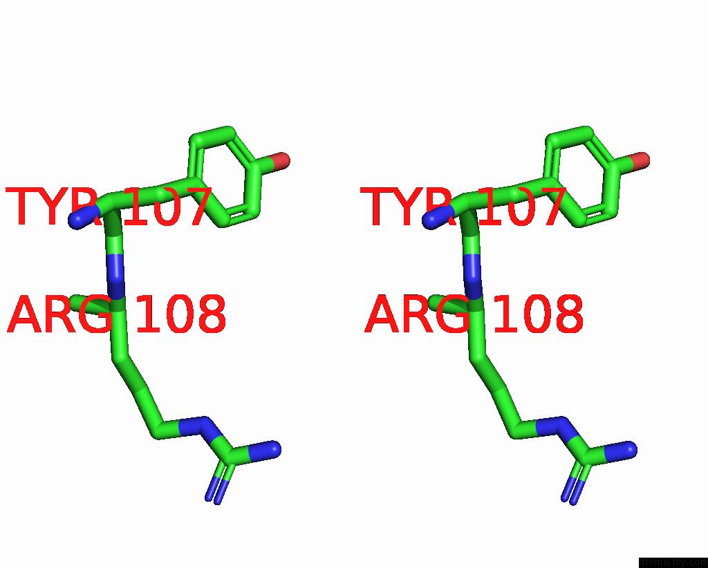
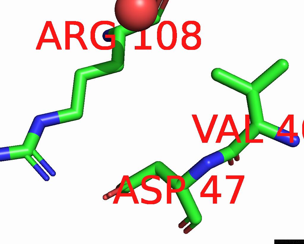
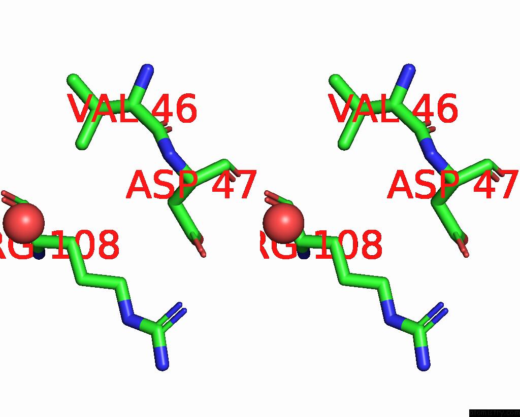
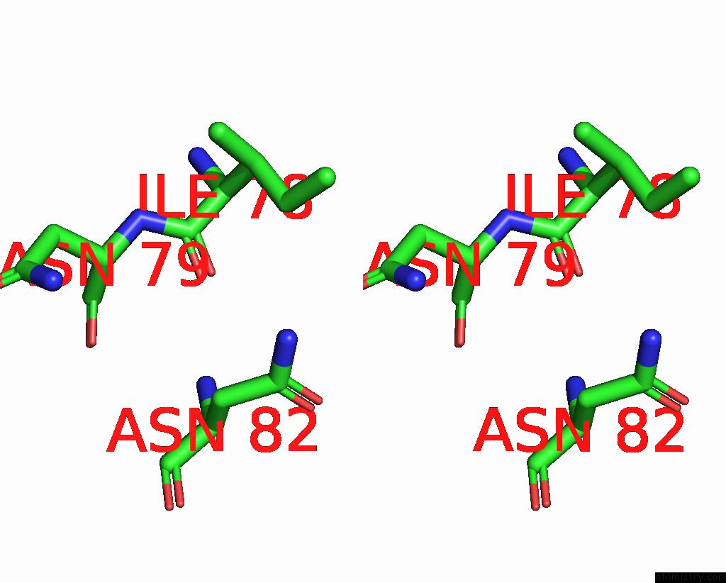
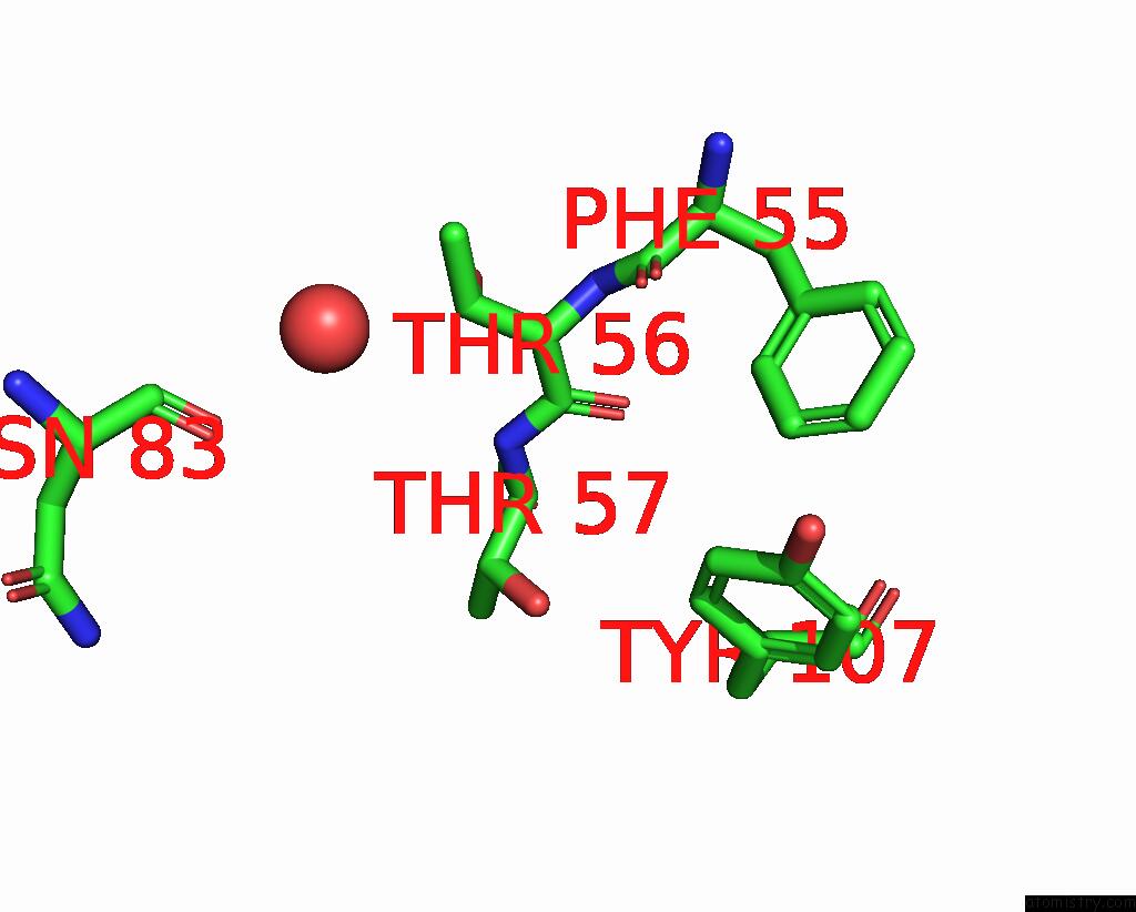
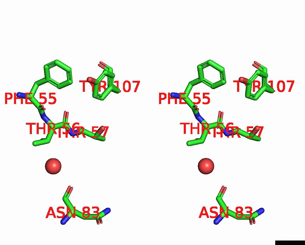
Iodine binding site 1 out of 12 in 8u0r
Go back to
Iodine binding site 1 out
of 12 in the The Crystal Structure of Protein A21, A Component of the Conserved Poxvirus Entry-Fusion Complex
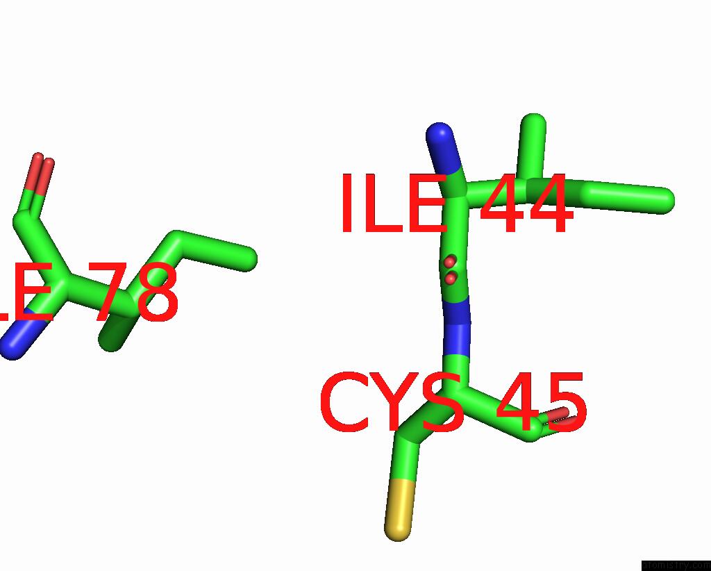
Mono view

Stereo pair view
Mono view
Stereo pair view
A full contact list of Iodine with other atoms in the I binding
site number 1 of The Crystal Structure of Protein A21, A Component of the Conserved Poxvirus Entry-Fusion Complex within 5.0Å range:
|
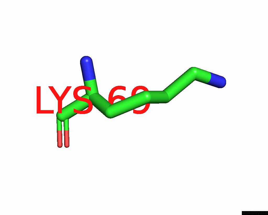
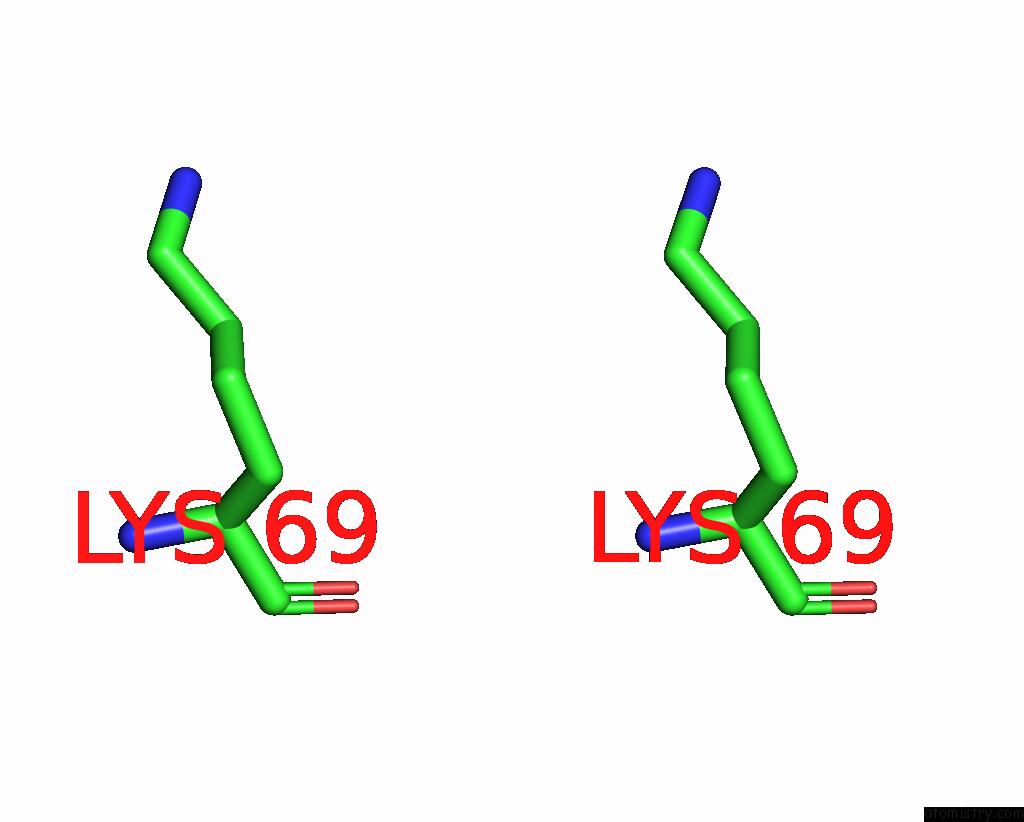
Iodine binding site 2 out of 12 in 8u0r
Go back to
Iodine binding site 2 out
of 12 in the The Crystal Structure of Protein A21, A Component of the Conserved Poxvirus Entry-Fusion Complex
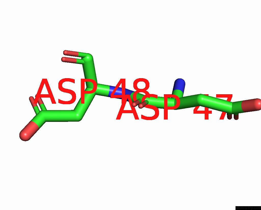
Mono view
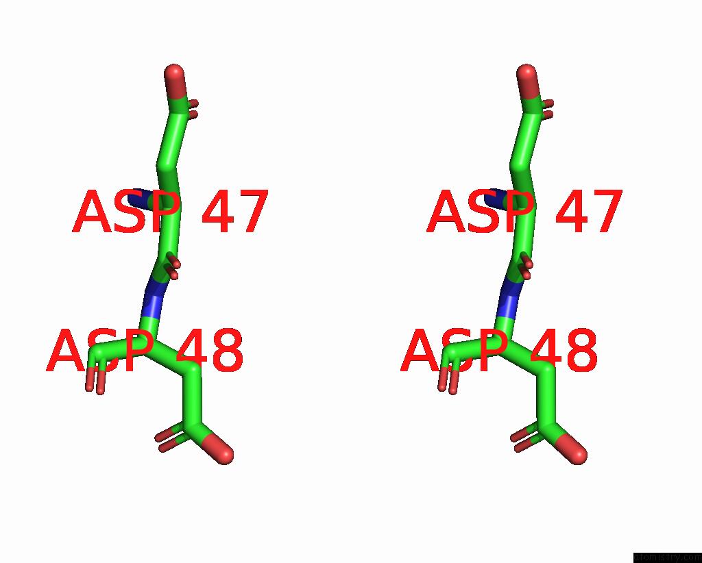
Stereo pair view
Mono view
Stereo pair view
A full contact list of Iodine with other atoms in the I binding
site number 2 of The Crystal Structure of Protein A21, A Component of the Conserved Poxvirus Entry-Fusion Complex within 5.0Å range:
|
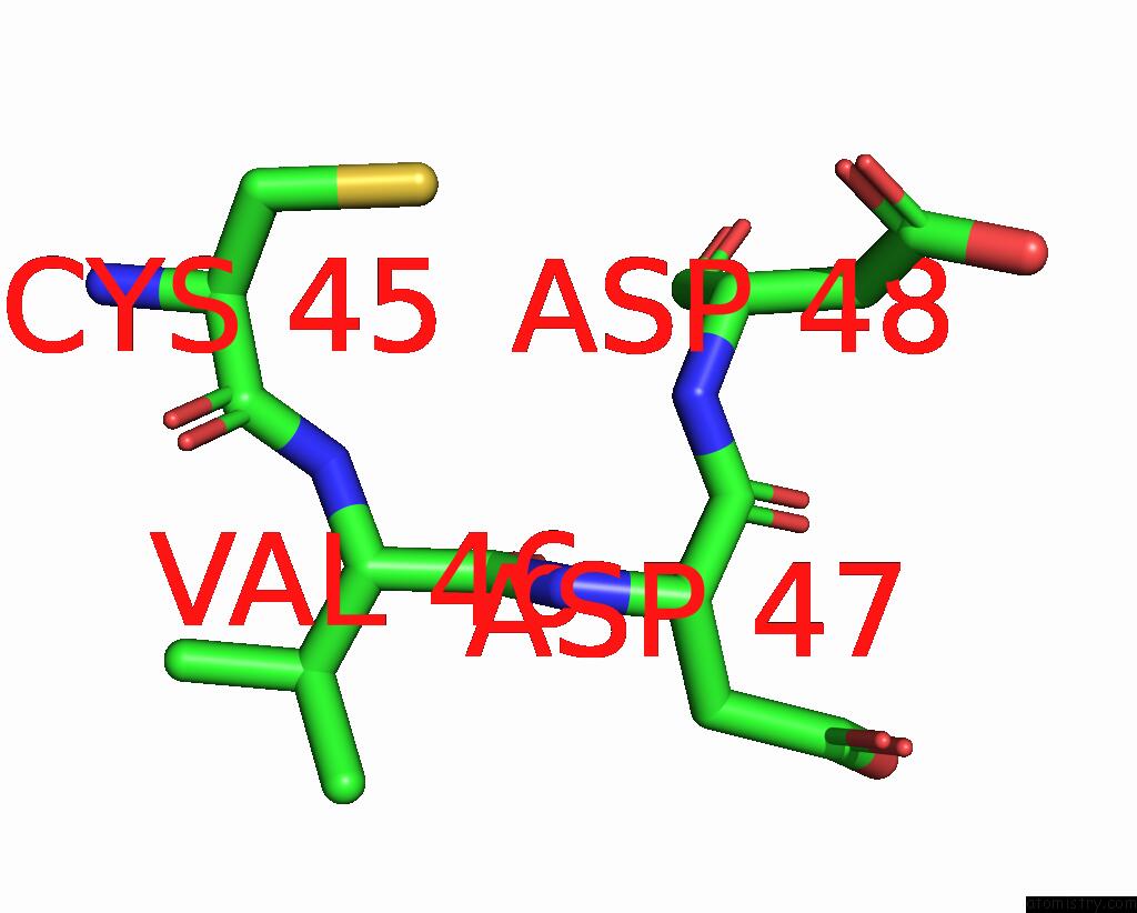
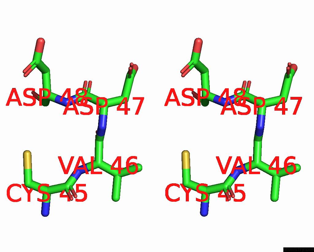
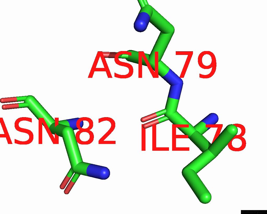
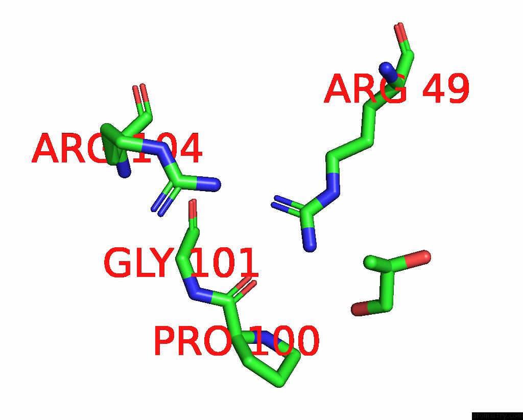
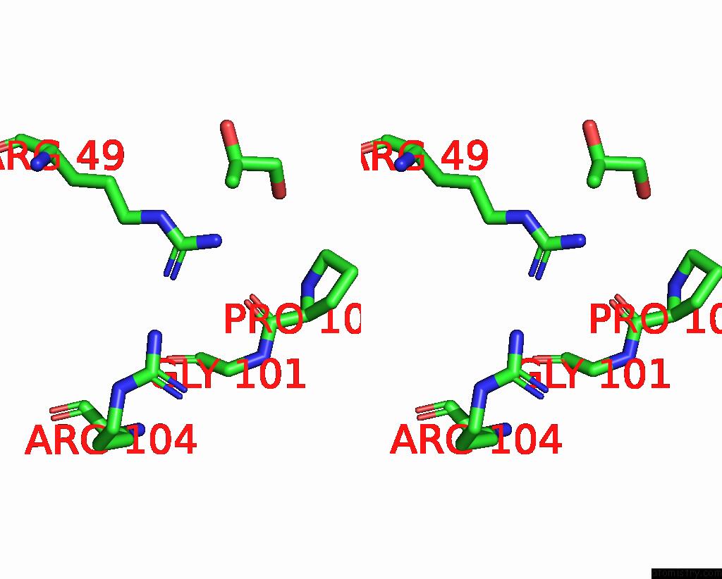
Iodine binding site 3 out of 12 in 8u0r
Go back to
Iodine binding site 3 out
of 12 in the The Crystal Structure of Protein A21, A Component of the Conserved Poxvirus Entry-Fusion Complex
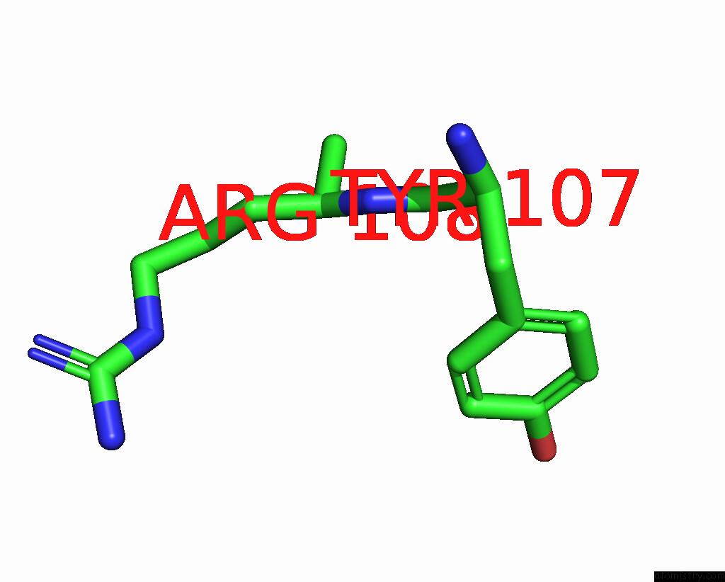
Mono view
Stereo pair view
Mono view
Stereo pair view
A full contact list of Iodine with other atoms in the I binding
site number 3 of The Crystal Structure of Protein A21, A Component of the Conserved Poxvirus Entry-Fusion Complex within 5.0Å range:
|
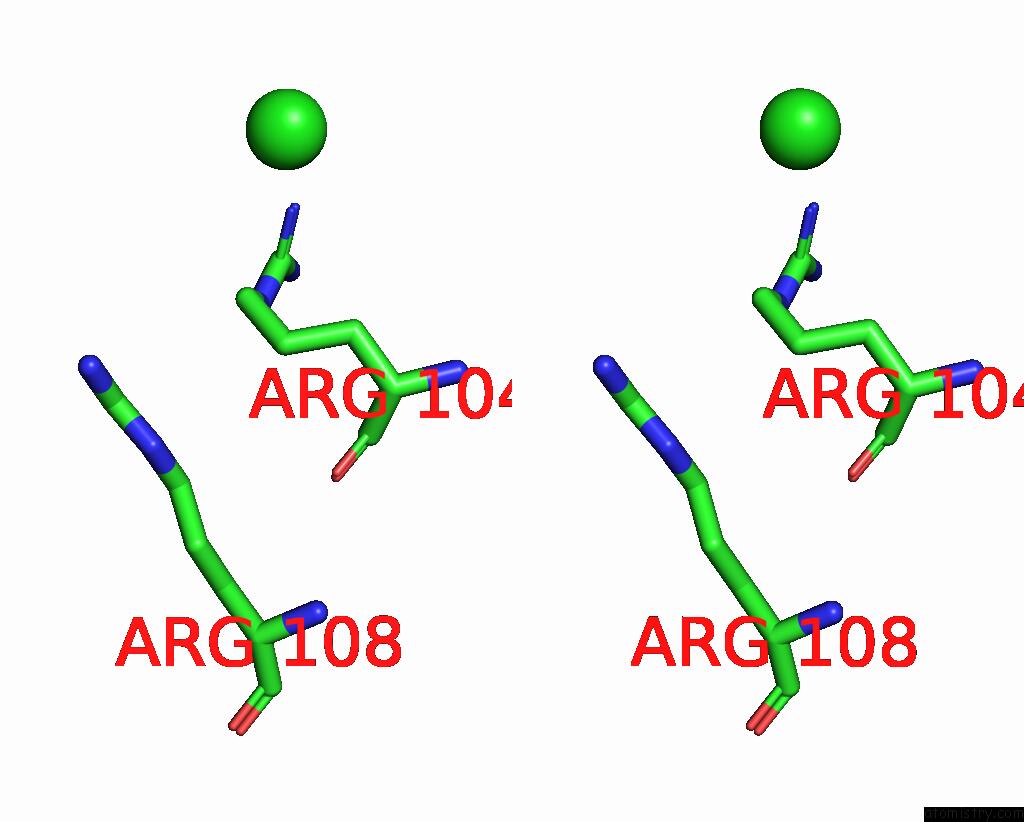
Iodine binding site 4 out of 12 in 8u0r
Go back to
Iodine binding site 4 out
of 12 in the The Crystal Structure of Protein A21, A Component of the Conserved Poxvirus Entry-Fusion Complex
Mono view
Stereo pair view
Mono view
Stereo pair view
A full contact list of Iodine with other atoms in the I binding
site number 4 of The Crystal Structure of Protein A21, A Component of the Conserved Poxvirus Entry-Fusion Complex within 5.0Å range:
|
Iodine binding site 5 out of 12 in 8u0r
Go back to
Iodine binding site 5 out
of 12 in the The Crystal Structure of Protein A21, A Component of the Conserved Poxvirus Entry-Fusion Complex
Mono view
Stereo pair view
Mono view
Stereo pair view
A full contact list of Iodine with other atoms in the I binding
site number 5 of The Crystal Structure of Protein A21, A Component of the Conserved Poxvirus Entry-Fusion Complex within 5.0Å range:
|
Iodine binding site 6 out of 12 in 8u0r
Go back to
Iodine binding site 6 out
of 12 in the The Crystal Structure of Protein A21, A Component of the Conserved Poxvirus Entry-Fusion Complex
Mono view
Stereo pair view
Mono view
Stereo pair view
A full contact list of Iodine with other atoms in the I binding
site number 6 of The Crystal Structure of Protein A21, A Component of the Conserved Poxvirus Entry-Fusion Complex within 5.0Å range:
|
Iodine binding site 7 out of 12 in 8u0r
Go back to
Iodine binding site 7 out
of 12 in the The Crystal Structure of Protein A21, A Component of the Conserved Poxvirus Entry-Fusion Complex
Mono view
Stereo pair view
Mono view
Stereo pair view
A full contact list of Iodine with other atoms in the I binding
site number 7 of The Crystal Structure of Protein A21, A Component of the Conserved Poxvirus Entry-Fusion Complex within 5.0Å range:
|
Iodine binding site 8 out of 12 in 8u0r
Go back to
Iodine binding site 8 out
of 12 in the The Crystal Structure of Protein A21, A Component of the Conserved Poxvirus Entry-Fusion Complex
Mono view
Stereo pair view
Mono view
Stereo pair view
A full contact list of Iodine with other atoms in the I binding
site number 8 of The Crystal Structure of Protein A21, A Component of the Conserved Poxvirus Entry-Fusion Complex within 5.0Å range:
|
Iodine binding site 9 out of 12 in 8u0r
Go back to
Iodine binding site 9 out
of 12 in the The Crystal Structure of Protein A21, A Component of the Conserved Poxvirus Entry-Fusion Complex

Mono view
Stereo pair view
Mono view
Stereo pair view
A full contact list of Iodine with other atoms in the I binding
site number 9 of The Crystal Structure of Protein A21, A Component of the Conserved Poxvirus Entry-Fusion Complex within 5.0Å range:
|
Iodine binding site 10 out of 12 in 8u0r
Go back to
Iodine binding site 10 out
of 12 in the The Crystal Structure of Protein A21, A Component of the Conserved Poxvirus Entry-Fusion Complex
Mono view
Stereo pair view
Mono view
Stereo pair view
A full contact list of Iodine with other atoms in the I binding
site number 10 of The Crystal Structure of Protein A21, A Component of the Conserved Poxvirus Entry-Fusion Complex within 5.0Å range:
|
Reference:
U.Diesterbeck,
A.Tak,
A.G.Gittis,
D.N.Garboczi,
B.Moss.
The Crystal Structure of Protein A21, A Component of the Conserved Poxvirus Entry-Fusion Complex To Be Published.
Page generated: Sat Sep 28 22:03:43 2024
Last articles
Cl in 2WSMCl in 2WT8
Cl in 2WSL
Cl in 2WSA
Cl in 2WSJ
Cl in 2WS7
Cl in 2WS6
Cl in 2WQ9
Cl in 2WQO
Cl in 2WR6