Iodine »
PDB 8ssv-9ku6 »
8ua2 »
Iodine in PDB 8ua2: Crystal Structure of Infected Cell Protein 0 (ICP0) From Herpes Simplex Virus 1 (Proteolyzed Fragment)
Protein crystallography data
The structure of Crystal Structure of Infected Cell Protein 0 (ICP0) From Herpes Simplex Virus 1 (Proteolyzed Fragment), PDB code: 8ua2
was solved by
S.Lovell,
M.Kashipathy,
K.P.Battaile,
A.Cooper,
D.Davido,
with X-Ray Crystallography technique. A brief refinement statistics is given in the table below:
| Resolution Low / High (Å) | 34.73 / 2.65 |
| Space group | P 41 21 2 |
| Cell size a, b, c (Å), α, β, γ (°) | 95.909, 95.909, 74.526, 90, 90, 90 |
| R / Rfree (%) | 21.8 / 28.8 |
Iodine Binding Sites:
The binding sites of Iodine atom in the Crystal Structure of Infected Cell Protein 0 (ICP0) From Herpes Simplex Virus 1 (Proteolyzed Fragment)
(pdb code 8ua2). This binding sites where shown within
5.0 Angstroms radius around Iodine atom.
In total 2 binding sites of Iodine where determined in the Crystal Structure of Infected Cell Protein 0 (ICP0) From Herpes Simplex Virus 1 (Proteolyzed Fragment), PDB code: 8ua2:
Jump to Iodine binding site number: 1; 2;
In total 2 binding sites of Iodine where determined in the Crystal Structure of Infected Cell Protein 0 (ICP0) From Herpes Simplex Virus 1 (Proteolyzed Fragment), PDB code: 8ua2:
Jump to Iodine binding site number: 1; 2;
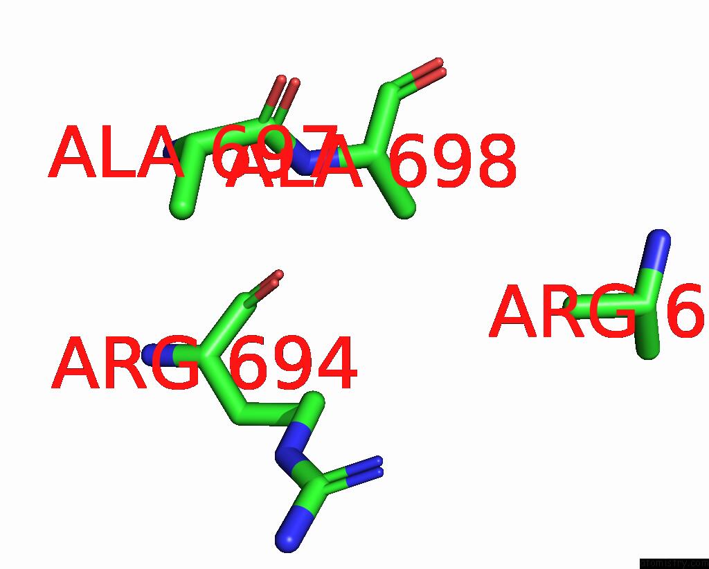
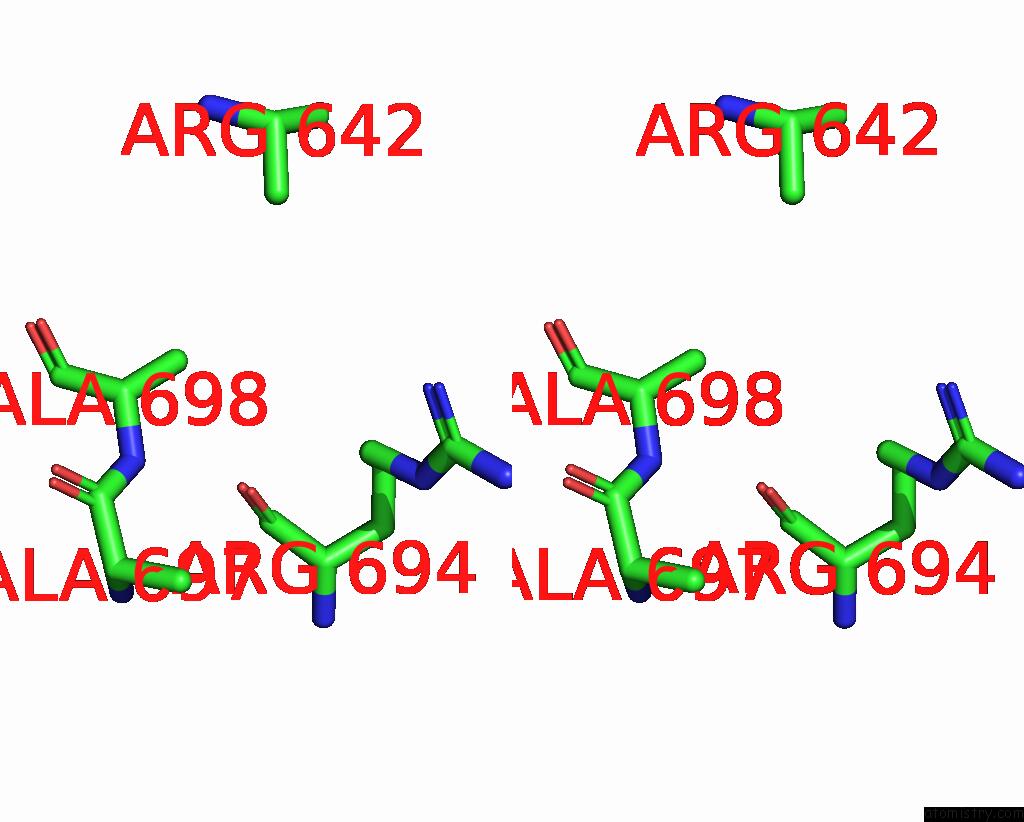
Iodine binding site 1 out of 2 in 8ua2
Go back to
Iodine binding site 1 out
of 2 in the Crystal Structure of Infected Cell Protein 0 (ICP0) From Herpes Simplex Virus 1 (Proteolyzed Fragment)
Mono view
Stereo pair view
Mono view
Stereo pair view
A full contact list of Iodine with other atoms in the I binding
site number 1 of Crystal Structure of Infected Cell Protein 0 (ICP0) From Herpes Simplex Virus 1 (Proteolyzed Fragment) within 5.0Å range:
|
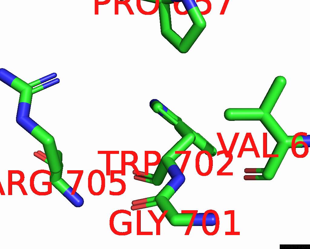
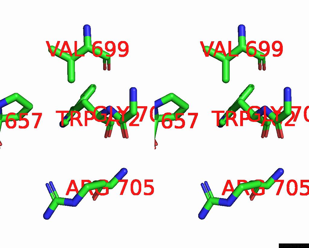
Iodine binding site 2 out of 2 in 8ua2
Go back to
Iodine binding site 2 out
of 2 in the Crystal Structure of Infected Cell Protein 0 (ICP0) From Herpes Simplex Virus 1 (Proteolyzed Fragment)
Mono view
Stereo pair view
Mono view
Stereo pair view
A full contact list of Iodine with other atoms in the I binding
site number 2 of Crystal Structure of Infected Cell Protein 0 (ICP0) From Herpes Simplex Virus 1 (Proteolyzed Fragment) within 5.0Å range:
|
Reference:
E.Mccloskey,
M.Kashipathy,
A.Cooper,
P.Gao,
D.K.Johnson,
K.P.Battaile,
S.Lovell,
D.J.Davido.
Hsv-1 ICP0 Dimer Domain Adopts A Novel Beta-Barrel Fold. Proteins 2024.
ISSN: ESSN 1097-0134
PubMed: 38372168
DOI: 10.1002/PROT.26673
Page generated: Mon Aug 12 03:11:36 2024
ISSN: ESSN 1097-0134
PubMed: 38372168
DOI: 10.1002/PROT.26673
Last articles
Zn in 9MJ5Zn in 9HNW
Zn in 9G0L
Zn in 9FNE
Zn in 9DZN
Zn in 9E0I
Zn in 9D32
Zn in 9DAK
Zn in 8ZXC
Zn in 8ZUF