Iodine »
PDB 8ssv-9ku6 »
9dq1 »
Iodine in PDB 9dq1: Crystal Structure of Hrmj From Streptomyces Sp. Cfmr 7 (Hrmj-Ssc) Complexed with Manganese (II), 2-Oxoglutarate and 6-Nitronorleucine
Protein crystallography data
The structure of Crystal Structure of Hrmj From Streptomyces Sp. Cfmr 7 (Hrmj-Ssc) Complexed with Manganese (II), 2-Oxoglutarate and 6-Nitronorleucine, PDB code: 9dq1
was solved by
Y.-C.Zheng,
P.Swartz,
W.-C.Chang,
with X-Ray Crystallography technique. A brief refinement statistics is given in the table below:
| Resolution Low / High (Å) | 17.49 / 1.90 |
| Space group | P 1 21 1 |
| Cell size a, b, c (Å), α, β, γ (°) | 44.342, 74.971, 87.295, 90, 90.03, 90 |
| R / Rfree (%) | 17.5 / 20.4 |
Other elements in 9dq1:
The structure of Crystal Structure of Hrmj From Streptomyces Sp. Cfmr 7 (Hrmj-Ssc) Complexed with Manganese (II), 2-Oxoglutarate and 6-Nitronorleucine also contains other interesting chemical elements:
| Manganese | (Mn) | 2 atoms |
Iodine Binding Sites:
The binding sites of Iodine atom in the Crystal Structure of Hrmj From Streptomyces Sp. Cfmr 7 (Hrmj-Ssc) Complexed with Manganese (II), 2-Oxoglutarate and 6-Nitronorleucine
(pdb code 9dq1). This binding sites where shown within
5.0 Angstroms radius around Iodine atom.
In total 7 binding sites of Iodine where determined in the Crystal Structure of Hrmj From Streptomyces Sp. Cfmr 7 (Hrmj-Ssc) Complexed with Manganese (II), 2-Oxoglutarate and 6-Nitronorleucine, PDB code: 9dq1:
Jump to Iodine binding site number: 1; 2; 3; 4; 5; 6; 7;
In total 7 binding sites of Iodine where determined in the Crystal Structure of Hrmj From Streptomyces Sp. Cfmr 7 (Hrmj-Ssc) Complexed with Manganese (II), 2-Oxoglutarate and 6-Nitronorleucine, PDB code: 9dq1:
Jump to Iodine binding site number: 1; 2; 3; 4; 5; 6; 7;
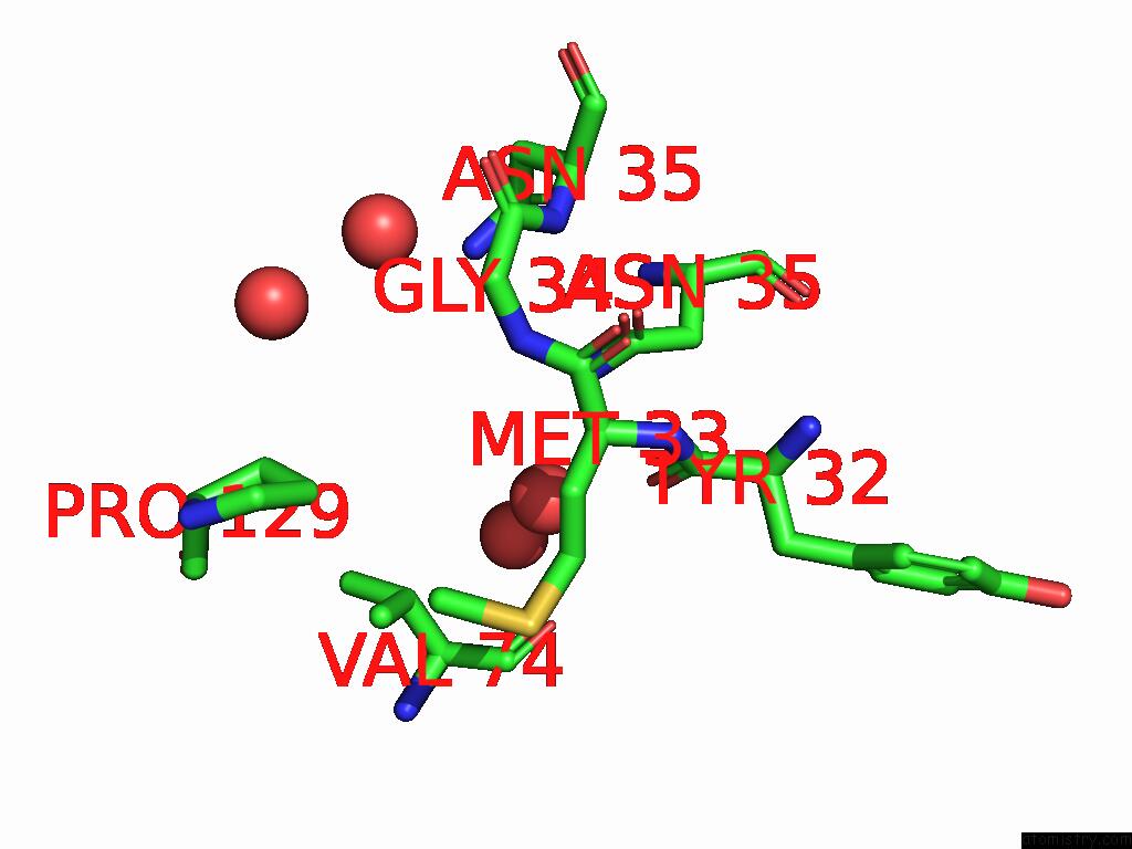
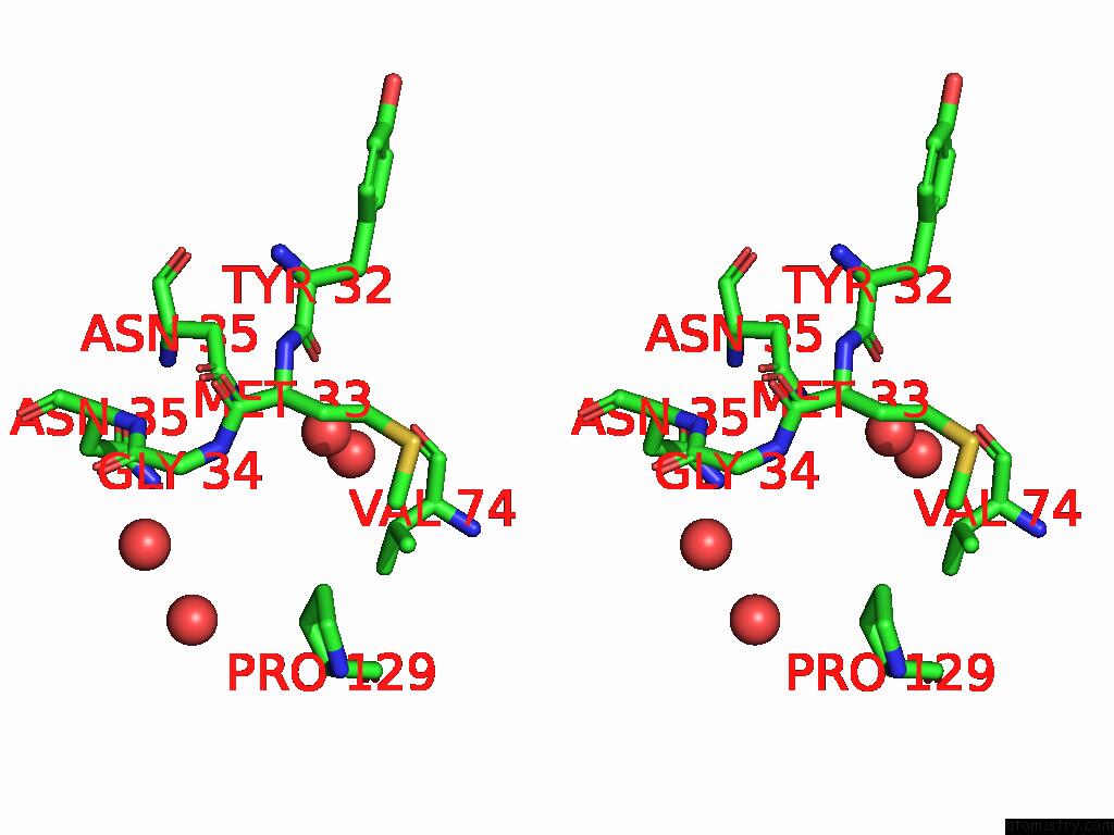
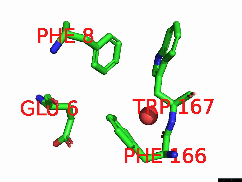
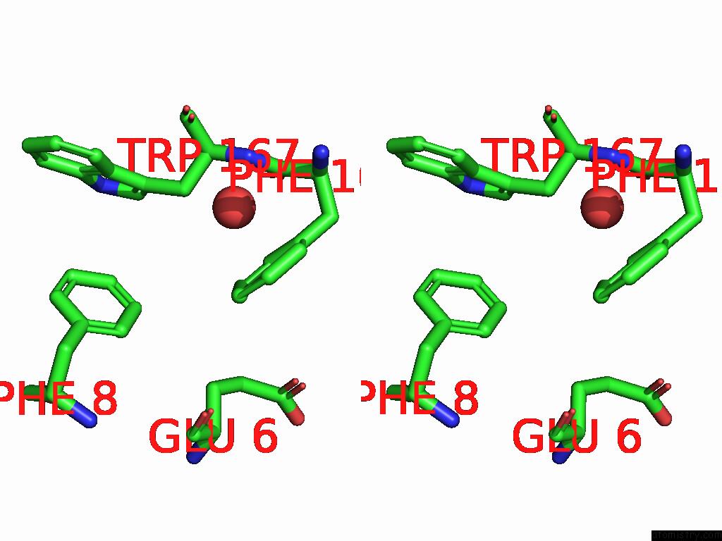
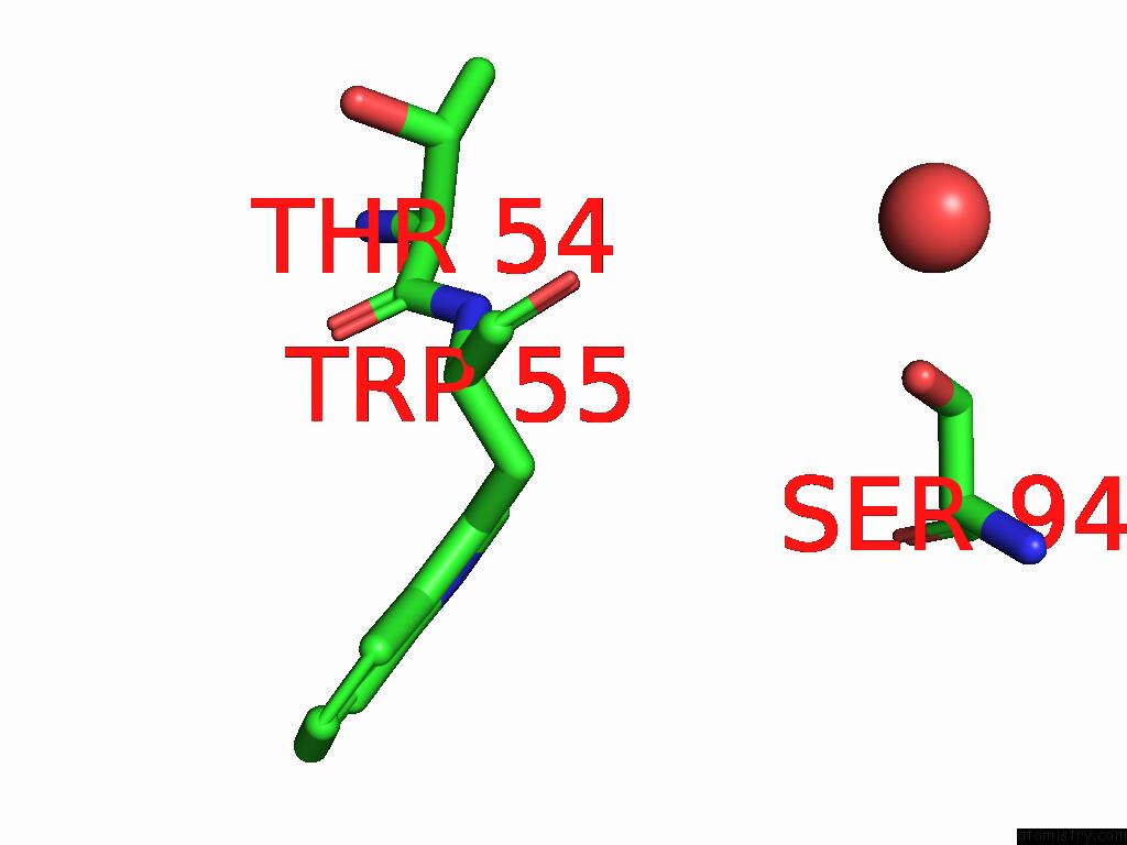
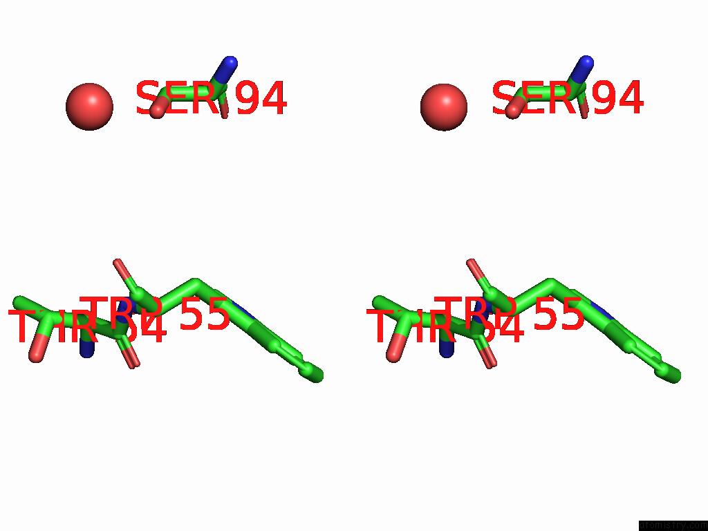
Iodine binding site 1 out of 7 in 9dq1
Go back to
Iodine binding site 1 out
of 7 in the Crystal Structure of Hrmj From Streptomyces Sp. Cfmr 7 (Hrmj-Ssc) Complexed with Manganese (II), 2-Oxoglutarate and 6-Nitronorleucine
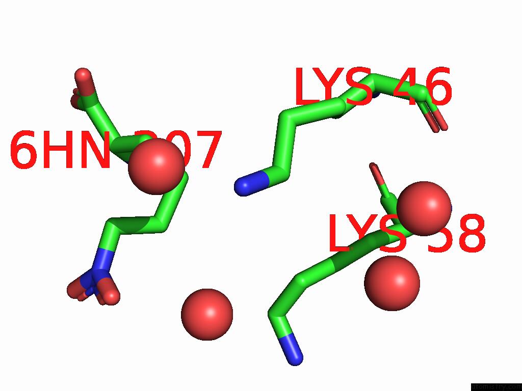
Mono view
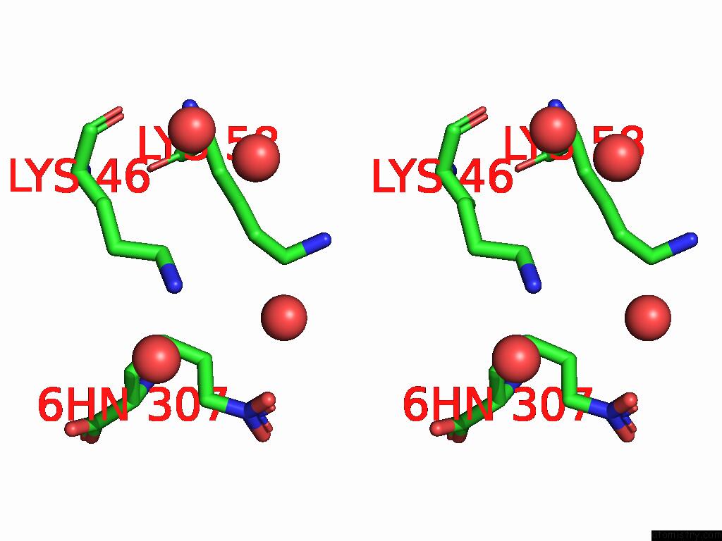
Stereo pair view
Mono view
Stereo pair view
A full contact list of Iodine with other atoms in the I binding
site number 1 of Crystal Structure of Hrmj From Streptomyces Sp. Cfmr 7 (Hrmj-Ssc) Complexed with Manganese (II), 2-Oxoglutarate and 6-Nitronorleucine within 5.0Å range:
|
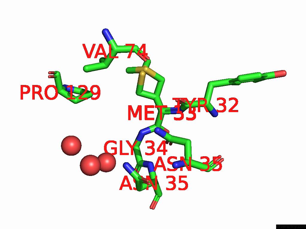
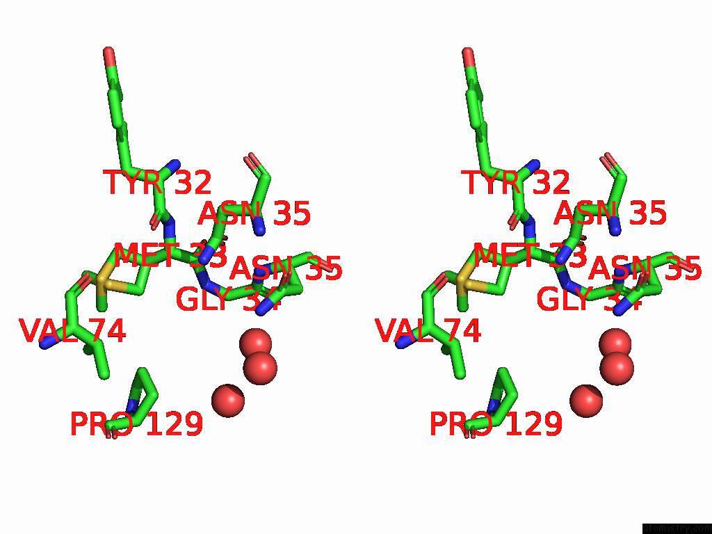
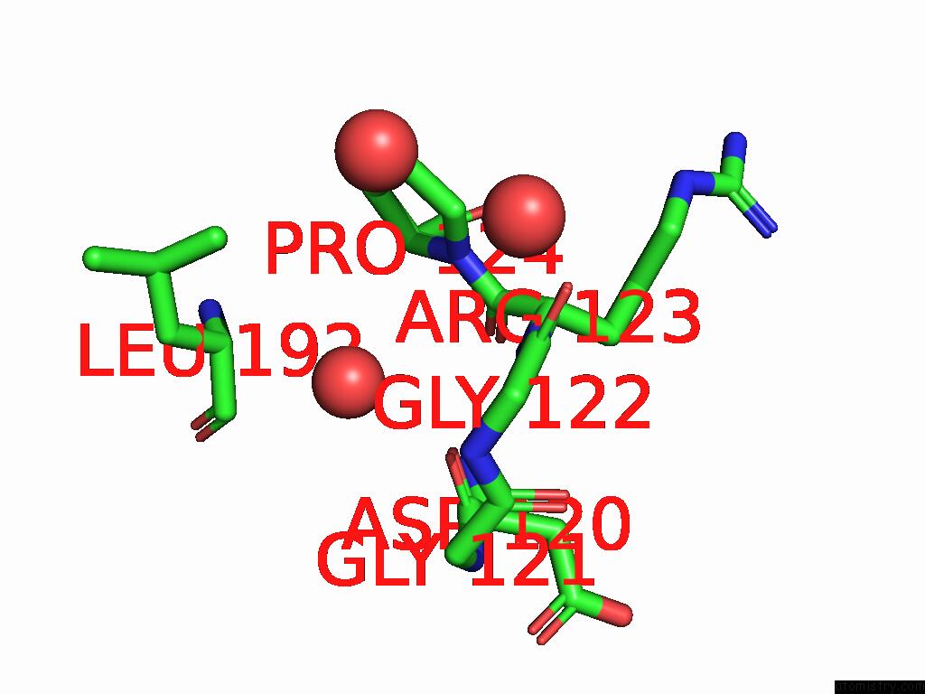
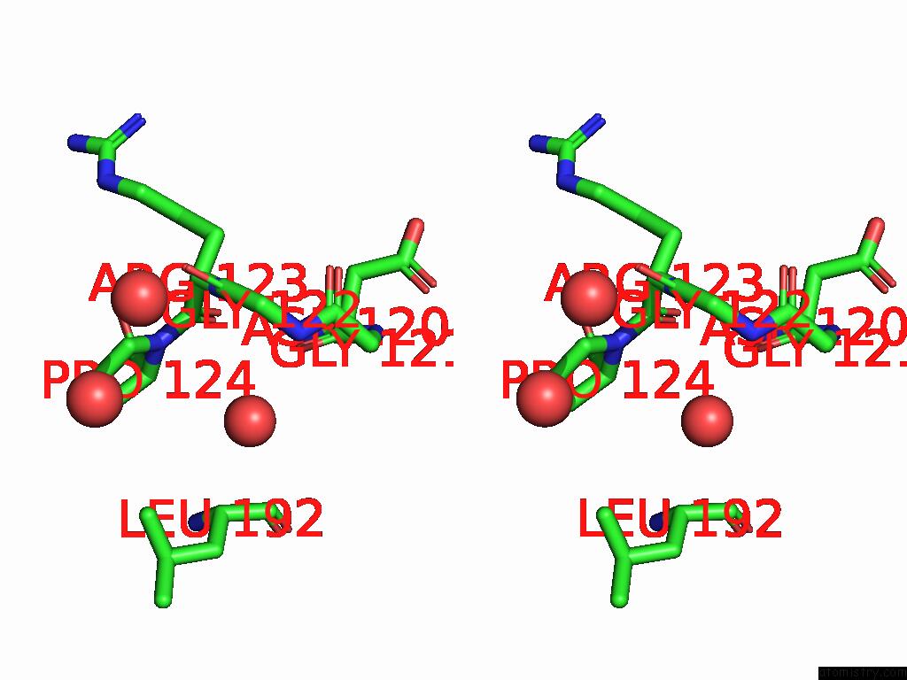
Iodine binding site 2 out of 7 in 9dq1
Go back to
Iodine binding site 2 out
of 7 in the Crystal Structure of Hrmj From Streptomyces Sp. Cfmr 7 (Hrmj-Ssc) Complexed with Manganese (II), 2-Oxoglutarate and 6-Nitronorleucine
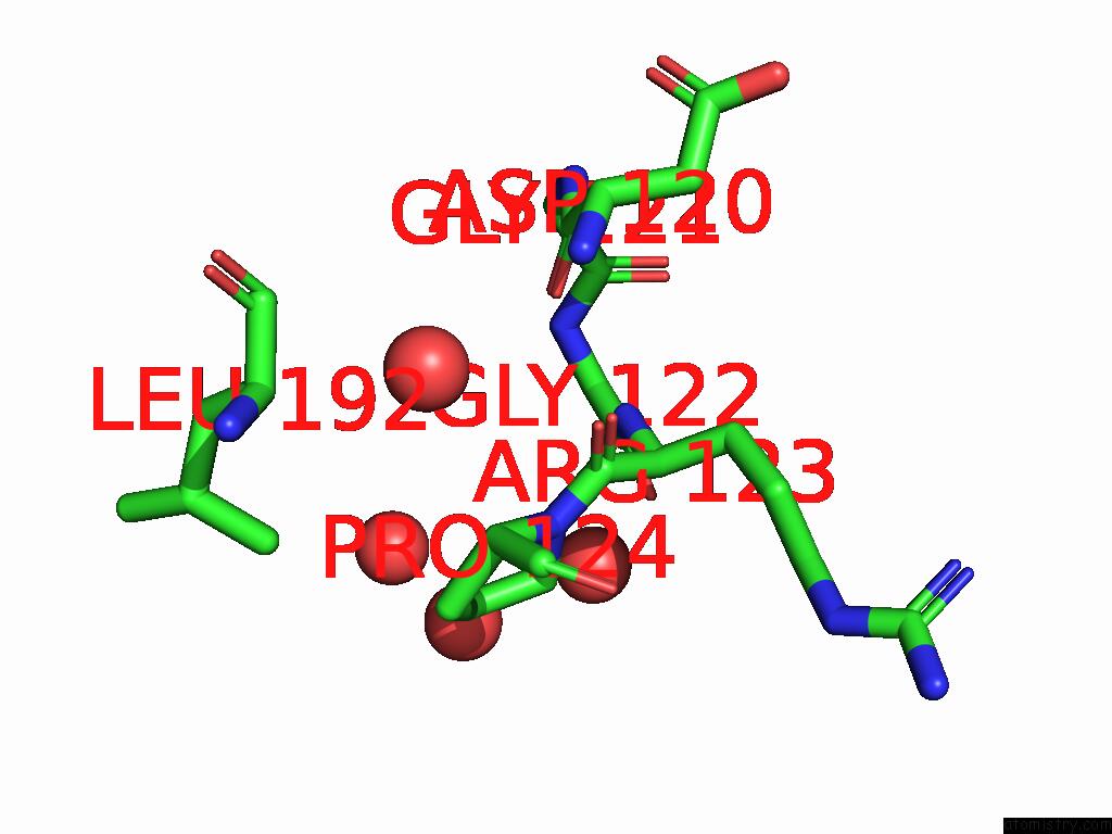
Mono view
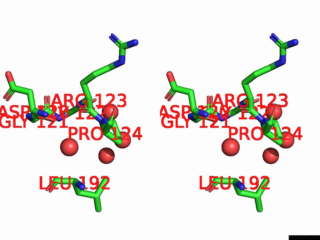
Stereo pair view
Mono view
Stereo pair view
A full contact list of Iodine with other atoms in the I binding
site number 2 of Crystal Structure of Hrmj From Streptomyces Sp. Cfmr 7 (Hrmj-Ssc) Complexed with Manganese (II), 2-Oxoglutarate and 6-Nitronorleucine within 5.0Å range:
|
Iodine binding site 3 out of 7 in 9dq1
Go back to
Iodine binding site 3 out
of 7 in the Crystal Structure of Hrmj From Streptomyces Sp. Cfmr 7 (Hrmj-Ssc) Complexed with Manganese (II), 2-Oxoglutarate and 6-Nitronorleucine
Mono view
Stereo pair view
Mono view
Stereo pair view
A full contact list of Iodine with other atoms in the I binding
site number 3 of Crystal Structure of Hrmj From Streptomyces Sp. Cfmr 7 (Hrmj-Ssc) Complexed with Manganese (II), 2-Oxoglutarate and 6-Nitronorleucine within 5.0Å range:
|
Iodine binding site 4 out of 7 in 9dq1
Go back to
Iodine binding site 4 out
of 7 in the Crystal Structure of Hrmj From Streptomyces Sp. Cfmr 7 (Hrmj-Ssc) Complexed with Manganese (II), 2-Oxoglutarate and 6-Nitronorleucine
Mono view
Stereo pair view
Mono view
Stereo pair view
A full contact list of Iodine with other atoms in the I binding
site number 4 of Crystal Structure of Hrmj From Streptomyces Sp. Cfmr 7 (Hrmj-Ssc) Complexed with Manganese (II), 2-Oxoglutarate and 6-Nitronorleucine within 5.0Å range:
|
Iodine binding site 5 out of 7 in 9dq1
Go back to
Iodine binding site 5 out
of 7 in the Crystal Structure of Hrmj From Streptomyces Sp. Cfmr 7 (Hrmj-Ssc) Complexed with Manganese (II), 2-Oxoglutarate and 6-Nitronorleucine
Mono view
Stereo pair view
Mono view
Stereo pair view
A full contact list of Iodine with other atoms in the I binding
site number 5 of Crystal Structure of Hrmj From Streptomyces Sp. Cfmr 7 (Hrmj-Ssc) Complexed with Manganese (II), 2-Oxoglutarate and 6-Nitronorleucine within 5.0Å range:
|
Iodine binding site 6 out of 7 in 9dq1
Go back to
Iodine binding site 6 out
of 7 in the Crystal Structure of Hrmj From Streptomyces Sp. Cfmr 7 (Hrmj-Ssc) Complexed with Manganese (II), 2-Oxoglutarate and 6-Nitronorleucine
Mono view
Stereo pair view
Mono view
Stereo pair view
A full contact list of Iodine with other atoms in the I binding
site number 6 of Crystal Structure of Hrmj From Streptomyces Sp. Cfmr 7 (Hrmj-Ssc) Complexed with Manganese (II), 2-Oxoglutarate and 6-Nitronorleucine within 5.0Å range:
|
Iodine binding site 7 out of 7 in 9dq1
Go back to
Iodine binding site 7 out
of 7 in the Crystal Structure of Hrmj From Streptomyces Sp. Cfmr 7 (Hrmj-Ssc) Complexed with Manganese (II), 2-Oxoglutarate and 6-Nitronorleucine
Mono view
Stereo pair view
Mono view
Stereo pair view
A full contact list of Iodine with other atoms in the I binding
site number 7 of Crystal Structure of Hrmj From Streptomyces Sp. Cfmr 7 (Hrmj-Ssc) Complexed with Manganese (II), 2-Oxoglutarate and 6-Nitronorleucine within 5.0Å range:
|
Reference:
Y.C.Zheng,
X.Li,
L.Cha,
J.C.Paris,
C.Michael,
R.Ushimaru,
Y.Ogasawara,
I.Abe,
Y.Guo,
W.C.Chang.
Comparison of A Nonheme Iron Cyclopropanase with A Homologous Hydroxylase Reveals Mechanistic Features Associated with Distinct Reaction Outcomes. J.Am.Chem.Soc. 2025.
ISSN: ESSN 1520-5126
PubMed: 39901767
DOI: 10.1021/JACS.4C17741
Page generated: Tue Feb 25 10:14:04 2025
ISSN: ESSN 1520-5126
PubMed: 39901767
DOI: 10.1021/JACS.4C17741
Last articles
Zn in 9MJ5Zn in 9HNW
Zn in 9G0L
Zn in 9FNE
Zn in 9DZN
Zn in 9E0I
Zn in 9D32
Zn in 9DAK
Zn in 8ZXC
Zn in 8ZUF