Iodine »
PDB 9axj-9rp9 »
9nwf »
Iodine in PDB 9nwf: Structure of An Inactive Beta-D-Glucuronate Dehydratase Mutant in Complex with Chondrosine
Protein crystallography data
The structure of Structure of An Inactive Beta-D-Glucuronate Dehydratase Mutant in Complex with Chondrosine, PDB code: 9nwf
was solved by
A.B.Boraston,
B.Alvarez,
O.Canil,
with X-Ray Crystallography technique. A brief refinement statistics is given in the table below:
| Resolution Low / High (Å) | 19.96 / 2.60 |
| Space group | P 21 21 21 |
| Cell size a, b, c (Å), α, β, γ (°) | 99.543, 105.84, 179.944, 90, 90, 90 |
| R / Rfree (%) | 19.1 / 26.2 |
Iodine Binding Sites:
Pages:
>>> Page 1 <<< Page 2, Binding sites: 11 - 20; Page 3, Binding sites: 21 - 27;Binding sites:
The binding sites of Iodine atom in the Structure of An Inactive Beta-D-Glucuronate Dehydratase Mutant in Complex with Chondrosine (pdb code 9nwf). This binding sites where shown within 5.0 Angstroms radius around Iodine atom.In total 27 binding sites of Iodine where determined in the Structure of An Inactive Beta-D-Glucuronate Dehydratase Mutant in Complex with Chondrosine, PDB code: 9nwf:
Jump to Iodine binding site number: 1; 2; 3; 4; 5; 6; 7; 8; 9; 10;
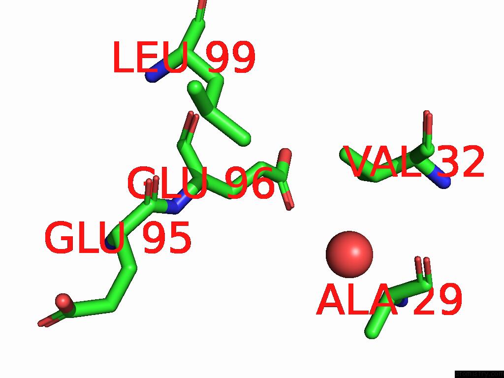
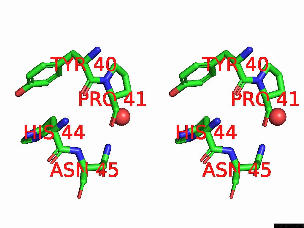
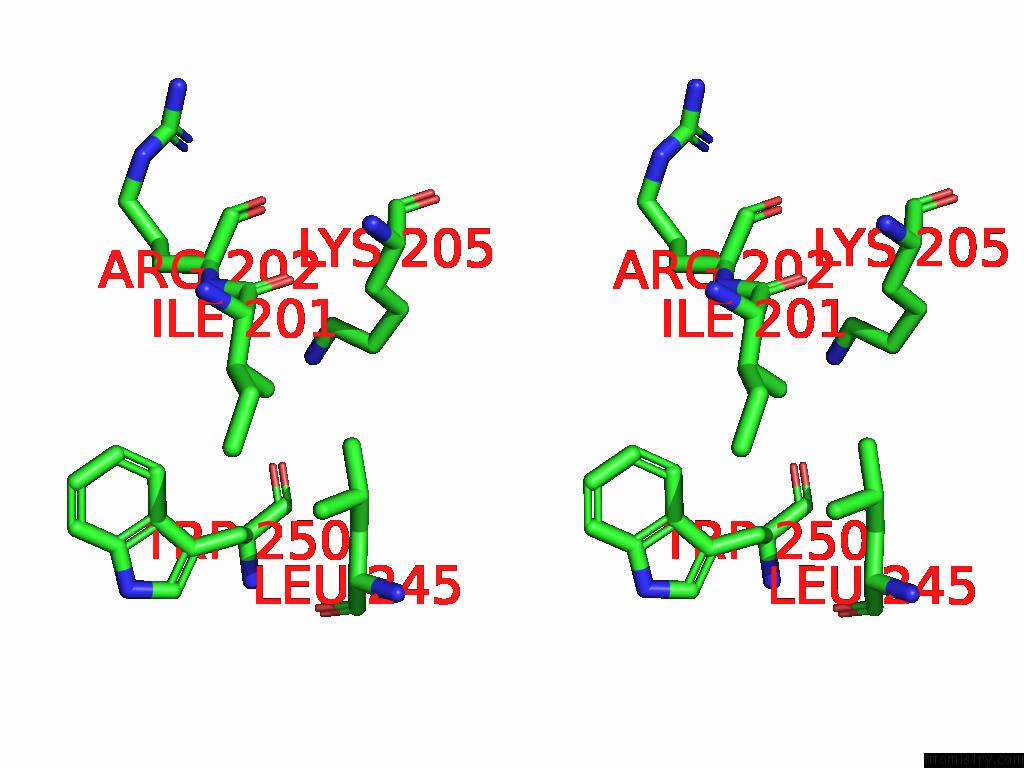
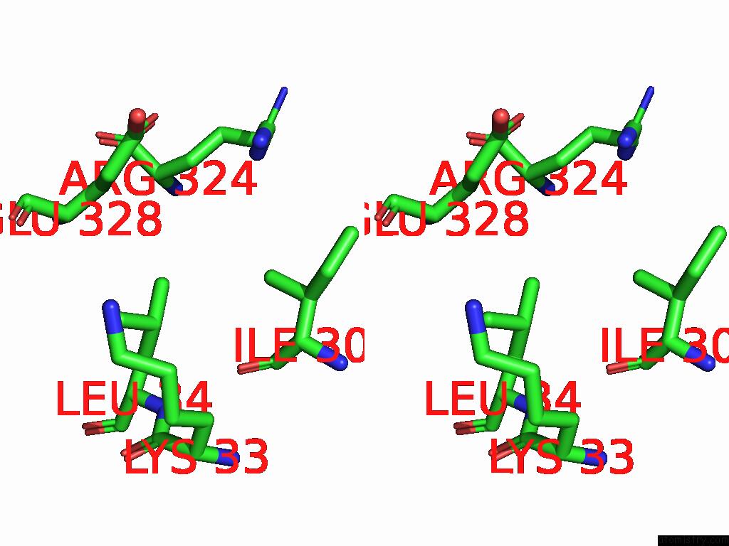
Iodine binding site 1 out of 27 in 9nwf
Go back to
Iodine binding site 1 out
of 27 in the Structure of An Inactive Beta-D-Glucuronate Dehydratase Mutant in Complex with Chondrosine
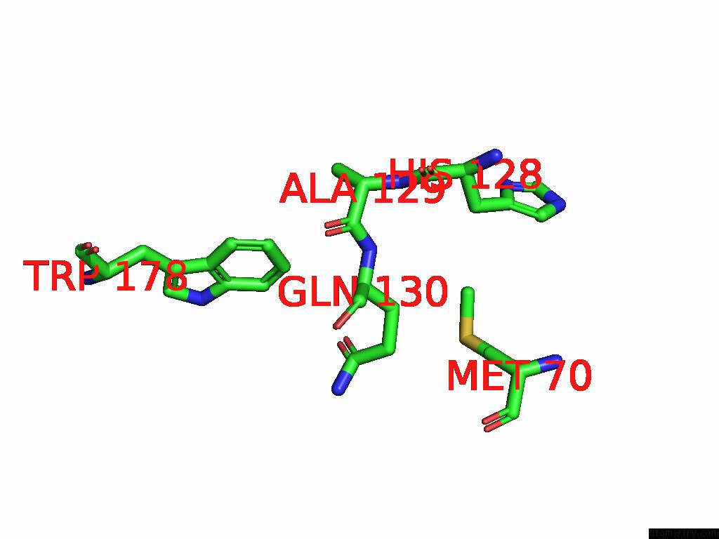
Mono view
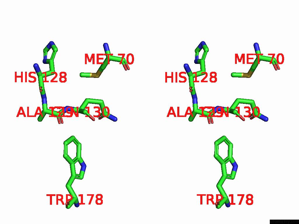
Stereo pair view
Mono view
Stereo pair view
A full contact list of Iodine with other atoms in the I binding
site number 1 of Structure of An Inactive Beta-D-Glucuronate Dehydratase Mutant in Complex with Chondrosine within 5.0Å range:
|
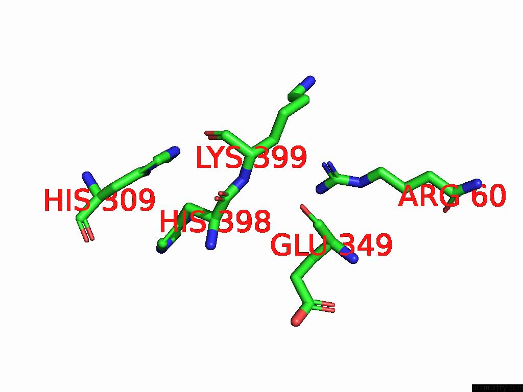
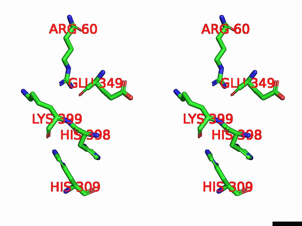
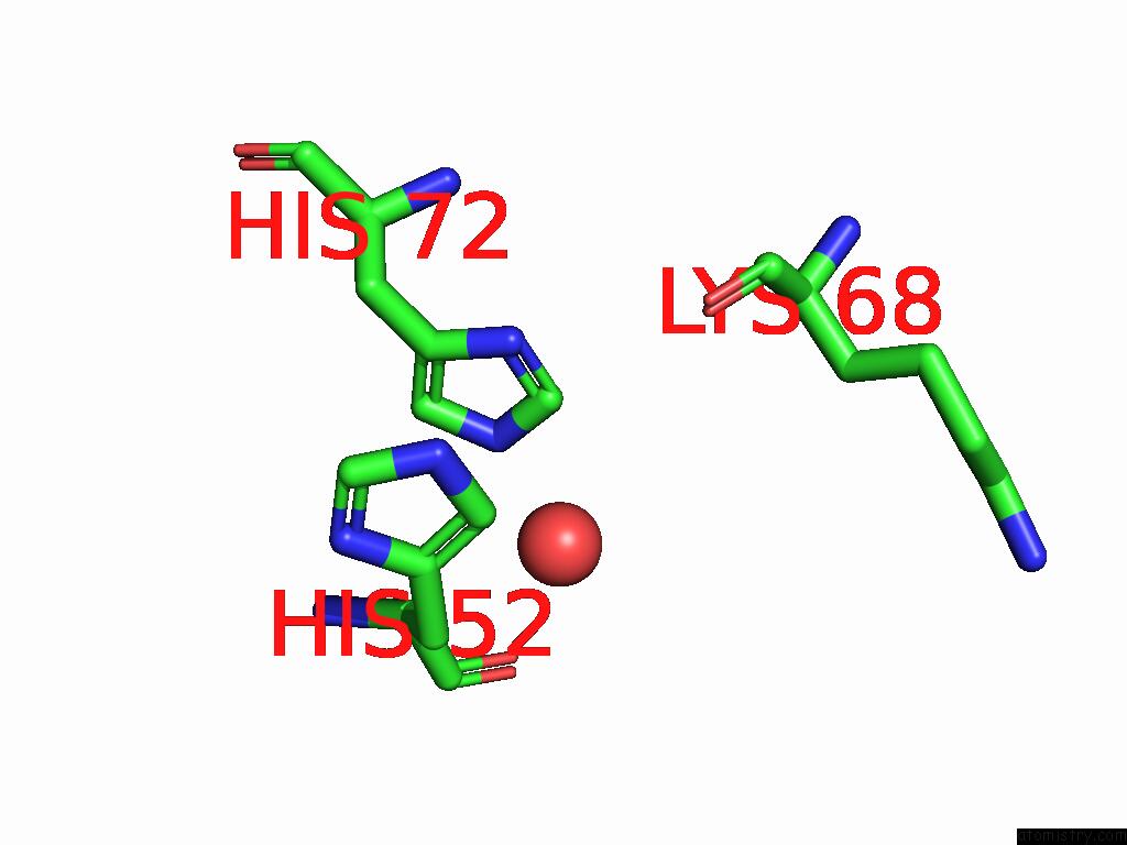
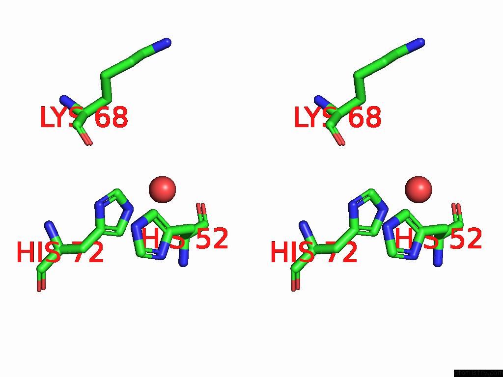
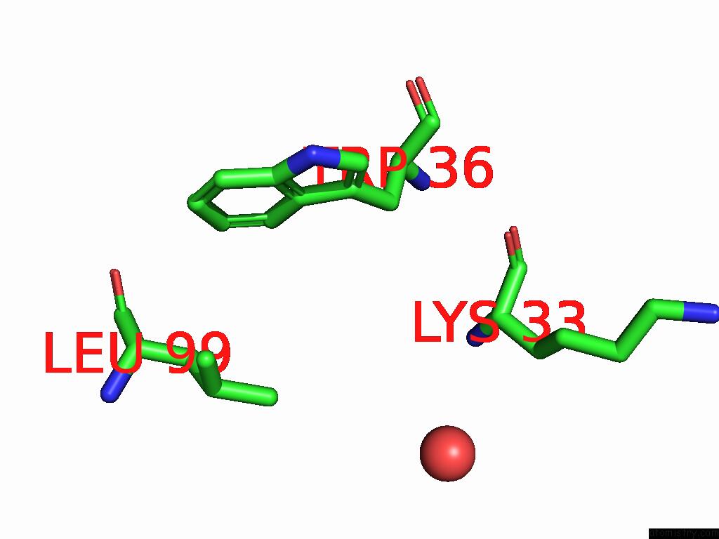
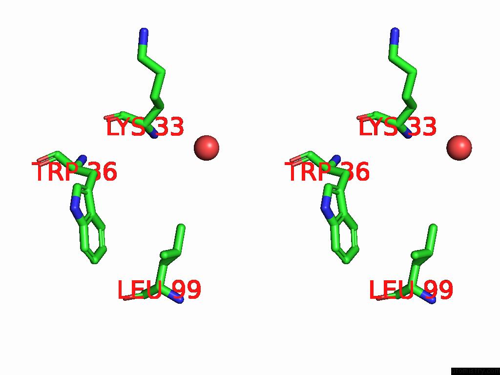
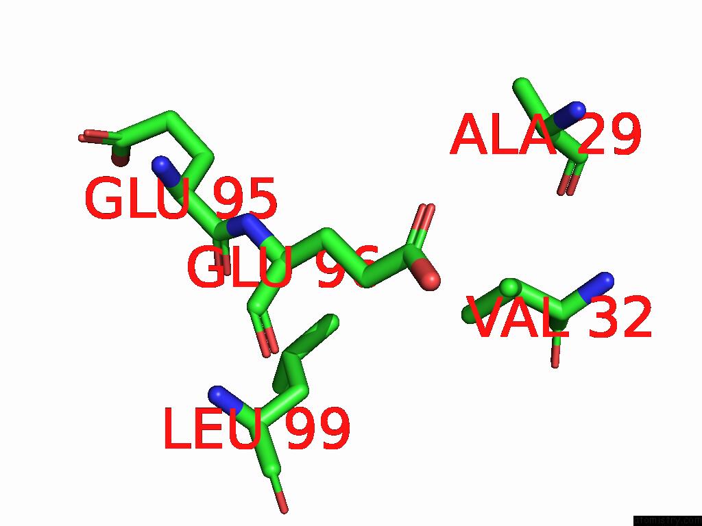
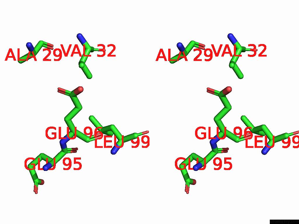
Iodine binding site 2 out of 27 in 9nwf
Go back to
Iodine binding site 2 out
of 27 in the Structure of An Inactive Beta-D-Glucuronate Dehydratase Mutant in Complex with Chondrosine
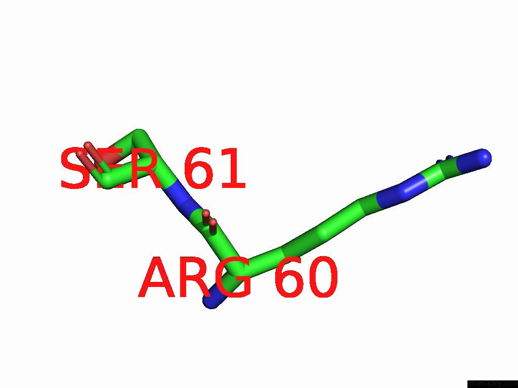
Mono view
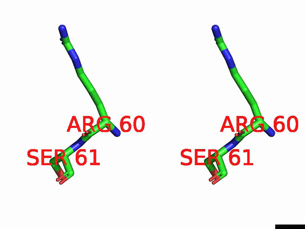
Stereo pair view
Mono view
Stereo pair view
A full contact list of Iodine with other atoms in the I binding
site number 2 of Structure of An Inactive Beta-D-Glucuronate Dehydratase Mutant in Complex with Chondrosine within 5.0Å range:
|
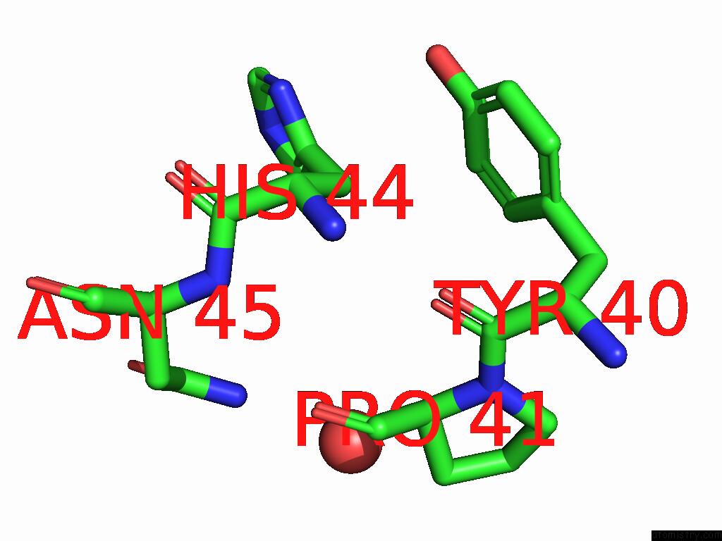
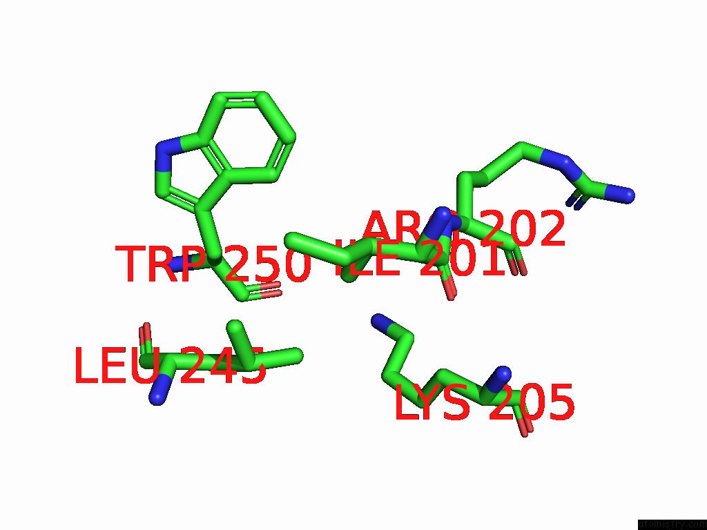
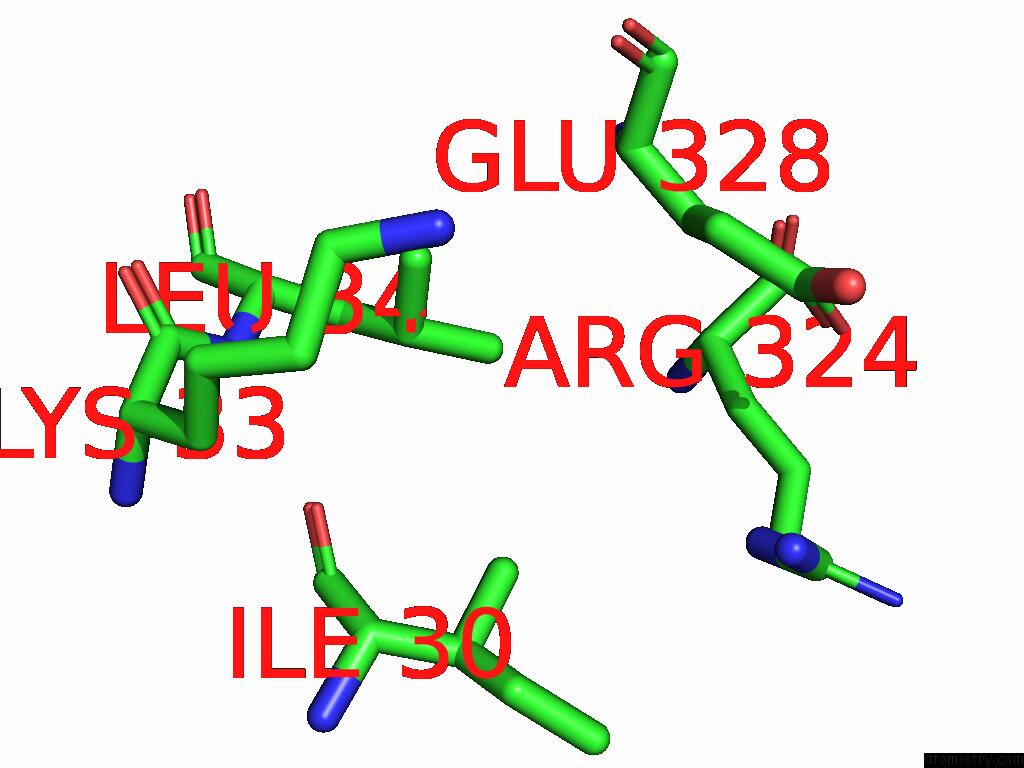
Iodine binding site 3 out of 27 in 9nwf
Go back to
Iodine binding site 3 out
of 27 in the Structure of An Inactive Beta-D-Glucuronate Dehydratase Mutant in Complex with Chondrosine
Mono view

Stereo pair view
Mono view
Stereo pair view
A full contact list of Iodine with other atoms in the I binding
site number 3 of Structure of An Inactive Beta-D-Glucuronate Dehydratase Mutant in Complex with Chondrosine within 5.0Å range:
|
Iodine binding site 4 out of 27 in 9nwf
Go back to
Iodine binding site 4 out
of 27 in the Structure of An Inactive Beta-D-Glucuronate Dehydratase Mutant in Complex with Chondrosine
Mono view
Stereo pair view
Mono view
Stereo pair view
A full contact list of Iodine with other atoms in the I binding
site number 4 of Structure of An Inactive Beta-D-Glucuronate Dehydratase Mutant in Complex with Chondrosine within 5.0Å range:
|
Iodine binding site 5 out of 27 in 9nwf
Go back to
Iodine binding site 5 out
of 27 in the Structure of An Inactive Beta-D-Glucuronate Dehydratase Mutant in Complex with Chondrosine
Mono view
Stereo pair view
Mono view
Stereo pair view
A full contact list of Iodine with other atoms in the I binding
site number 5 of Structure of An Inactive Beta-D-Glucuronate Dehydratase Mutant in Complex with Chondrosine within 5.0Å range:
|
Iodine binding site 6 out of 27 in 9nwf
Go back to
Iodine binding site 6 out
of 27 in the Structure of An Inactive Beta-D-Glucuronate Dehydratase Mutant in Complex with Chondrosine
Mono view
Stereo pair view
Mono view
Stereo pair view
A full contact list of Iodine with other atoms in the I binding
site number 6 of Structure of An Inactive Beta-D-Glucuronate Dehydratase Mutant in Complex with Chondrosine within 5.0Å range:
|
Iodine binding site 7 out of 27 in 9nwf
Go back to
Iodine binding site 7 out
of 27 in the Structure of An Inactive Beta-D-Glucuronate Dehydratase Mutant in Complex with Chondrosine
Mono view
Stereo pair view
Mono view
Stereo pair view
A full contact list of Iodine with other atoms in the I binding
site number 7 of Structure of An Inactive Beta-D-Glucuronate Dehydratase Mutant in Complex with Chondrosine within 5.0Å range:
|
Iodine binding site 8 out of 27 in 9nwf
Go back to
Iodine binding site 8 out
of 27 in the Structure of An Inactive Beta-D-Glucuronate Dehydratase Mutant in Complex with Chondrosine
Mono view
Stereo pair view
Mono view
Stereo pair view
A full contact list of Iodine with other atoms in the I binding
site number 8 of Structure of An Inactive Beta-D-Glucuronate Dehydratase Mutant in Complex with Chondrosine within 5.0Å range:
|
Iodine binding site 9 out of 27 in 9nwf
Go back to
Iodine binding site 9 out
of 27 in the Structure of An Inactive Beta-D-Glucuronate Dehydratase Mutant in Complex with Chondrosine
Mono view
Stereo pair view
Mono view
Stereo pair view
A full contact list of Iodine with other atoms in the I binding
site number 9 of Structure of An Inactive Beta-D-Glucuronate Dehydratase Mutant in Complex with Chondrosine within 5.0Å range:
|
Iodine binding site 10 out of 27 in 9nwf
Go back to
Iodine binding site 10 out
of 27 in the Structure of An Inactive Beta-D-Glucuronate Dehydratase Mutant in Complex with Chondrosine
Mono view
Stereo pair view
Mono view
Stereo pair view
A full contact list of Iodine with other atoms in the I binding
site number 10 of Structure of An Inactive Beta-D-Glucuronate Dehydratase Mutant in Complex with Chondrosine within 5.0Å range:
|
Reference:
B.Alvarez,
O.Canil,
K.Low,
D.W.Abbott,
A.B.Boraston.
Analysis of Chondroitin Degradation By Components of A Bacteroides Caccae Polysaccharide Utilization Locus To Be Published.
Page generated: Sat Aug 9 00:53:35 2025
Last articles
K in 3LLPK in 3LMS
K in 3LM7
K in 3LDD
K in 3LDC
K in 3LKI
K in 3L01
K in 3L63
K in 3KTW
K in 3L8D