Iodine »
PDB 1lkr-1pvh »
1mqg »
Iodine in PDB 1mqg: Crystal Structure of the GLUR2 Ligand Binding Core (S1S2J) in Complex with Iodo-Willardiine at 2.15 Angstroms Resolution
Protein crystallography data
The structure of Crystal Structure of the GLUR2 Ligand Binding Core (S1S2J) in Complex with Iodo-Willardiine at 2.15 Angstroms Resolution, PDB code: 1mqg
was solved by
R.Jin,
T.G.Banke,
M.L.Mayer,
S.F.Traynelis,
E.Gouaux,
with X-Ray Crystallography technique. A brief refinement statistics is given in the table below:
| Resolution Low / High (Å) | 29.71 / 2.15 |
| Space group | P 1 21 1 |
| Cell size a, b, c (Å), α, β, γ (°) | 48.006, 88.716, 58.907, 90.00, 99.24, 90.00 |
| R / Rfree (%) | 18.5 / 23.3 |
Iodine Binding Sites:
The binding sites of Iodine atom in the Crystal Structure of the GLUR2 Ligand Binding Core (S1S2J) in Complex with Iodo-Willardiine at 2.15 Angstroms Resolution
(pdb code 1mqg). This binding sites where shown within
5.0 Angstroms radius around Iodine atom.
In total 3 binding sites of Iodine where determined in the Crystal Structure of the GLUR2 Ligand Binding Core (S1S2J) in Complex with Iodo-Willardiine at 2.15 Angstroms Resolution, PDB code: 1mqg:
Jump to Iodine binding site number: 1; 2; 3;
In total 3 binding sites of Iodine where determined in the Crystal Structure of the GLUR2 Ligand Binding Core (S1S2J) in Complex with Iodo-Willardiine at 2.15 Angstroms Resolution, PDB code: 1mqg:
Jump to Iodine binding site number: 1; 2; 3;
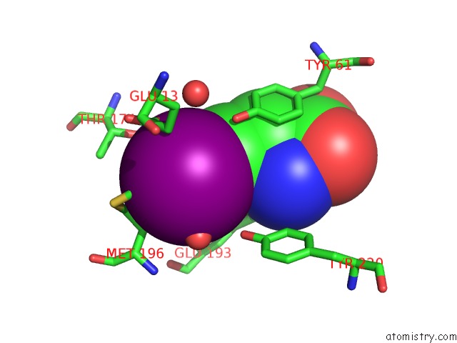
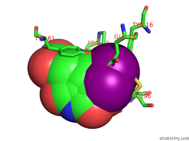
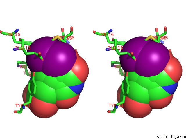
Iodine binding site 1 out of 3 in 1mqg
Go back to
Iodine binding site 1 out
of 3 in the Crystal Structure of the GLUR2 Ligand Binding Core (S1S2J) in Complex with Iodo-Willardiine at 2.15 Angstroms Resolution
Mono view
Stereo pair view
Mono view
Stereo pair view
|
|
A full contact list of Iodine with other atoms in the I binding
site number 1 of Crystal Structure of the GLUR2 Ligand Binding Core (S1S2J) in Complex with Iodo-Willardiine at 2.15 Angstroms Resolution within 5.0Å range:
|
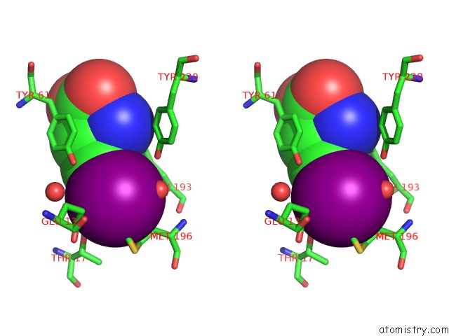
Iodine binding site 2 out of 3 in 1mqg
Go back to
Iodine binding site 2 out
of 3 in the Crystal Structure of the GLUR2 Ligand Binding Core (S1S2J) in Complex with Iodo-Willardiine at 2.15 Angstroms Resolution
Mono view
Stereo pair view
Mono view
Stereo pair view
|
|
A full contact list of Iodine with other atoms in the I binding
site number 2 of Crystal Structure of the GLUR2 Ligand Binding Core (S1S2J) in Complex with Iodo-Willardiine at 2.15 Angstroms Resolution within 5.0Å range:
|
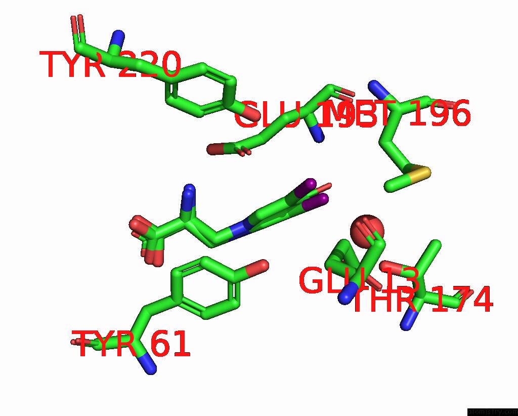
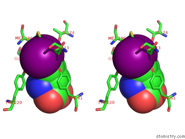
Iodine binding site 3 out of 3 in 1mqg
Go back to
Iodine binding site 3 out
of 3 in the Crystal Structure of the GLUR2 Ligand Binding Core (S1S2J) in Complex with Iodo-Willardiine at 2.15 Angstroms Resolution
Mono view
Stereo pair view
Mono view
Stereo pair view
|
|
A full contact list of Iodine with other atoms in the I binding
site number 3 of Crystal Structure of the GLUR2 Ligand Binding Core (S1S2J) in Complex with Iodo-Willardiine at 2.15 Angstroms Resolution within 5.0Å range:
|
Reference:
R.Jin,
T.G.Banke,
M.L.Mayer,
S.F.Traynelis,
E.Gouaux.
Structural Basis For Partial Agonist Action at Ionotropic Glutamate Receptors Nat.Neurosci. V. 6 803 2003.
ISSN: ISSN 1097-6256
PubMed: 12872125
DOI: 10.1038/NN1091
Page generated: Sun Aug 11 12:28:47 2024
ISSN: ISSN 1097-6256
PubMed: 12872125
DOI: 10.1038/NN1091
Last articles
Fe in 6CDKFe in 6CII
Fe in 6CFW
Fe in 6CIF
Fe in 6CIE
Fe in 6CID
Fe in 6CIC
Fe in 6CHI
Fe in 6CI0
Fe in 6CH6