Iodine »
PDB 1lkr-1pvh »
1orw »
Iodine in PDB 1orw: Crystal Structure of Porcine Dipeptidyl Peptidase IV (CD26) in Complex with A Peptidomimetic Inhibitor
Enzymatic activity of Crystal Structure of Porcine Dipeptidyl Peptidase IV (CD26) in Complex with A Peptidomimetic Inhibitor
All present enzymatic activity of Crystal Structure of Porcine Dipeptidyl Peptidase IV (CD26) in Complex with A Peptidomimetic Inhibitor:
3.4.14.5;
3.4.14.5;
Protein crystallography data
The structure of Crystal Structure of Porcine Dipeptidyl Peptidase IV (CD26) in Complex with A Peptidomimetic Inhibitor, PDB code: 1orw
was solved by
M.Engel,
T.Hoffmann,
L.Wagner,
M.Wermann,
U.Heiser,
R.Kiefersauer,
R.Huber,
W.Bode,
H.U.Demuth,
H.Brandstetter,
with X-Ray Crystallography technique. A brief refinement statistics is given in the table below:
| Resolution Low / High (Å) | 29.79 / 2.84 |
| Space group | P 1 |
| Cell size a, b, c (Å), α, β, γ (°) | 61.970, 117.710, 133.560, 112.65, 94.81, 91.17 |
| R / Rfree (%) | 18.7 / 24.6 |
Iodine Binding Sites:
The binding sites of Iodine atom in the Crystal Structure of Porcine Dipeptidyl Peptidase IV (CD26) in Complex with A Peptidomimetic Inhibitor
(pdb code 1orw). This binding sites where shown within
5.0 Angstroms radius around Iodine atom.
In total 4 binding sites of Iodine where determined in the Crystal Structure of Porcine Dipeptidyl Peptidase IV (CD26) in Complex with A Peptidomimetic Inhibitor, PDB code: 1orw:
Jump to Iodine binding site number: 1; 2; 3; 4;
In total 4 binding sites of Iodine where determined in the Crystal Structure of Porcine Dipeptidyl Peptidase IV (CD26) in Complex with A Peptidomimetic Inhibitor, PDB code: 1orw:
Jump to Iodine binding site number: 1; 2; 3; 4;
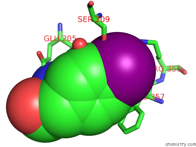
Iodine binding site 1 out of 4 in 1orw
Go back to
Iodine binding site 1 out
of 4 in the Crystal Structure of Porcine Dipeptidyl Peptidase IV (CD26) in Complex with A Peptidomimetic Inhibitor
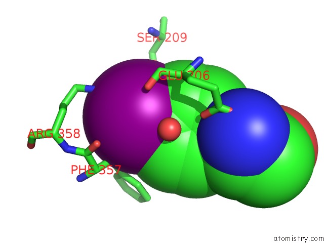
Mono view
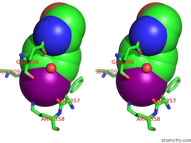
Stereo pair view
Mono view
Stereo pair view
A full contact list of Iodine with other atoms in the I binding
site number 1 of Crystal Structure of Porcine Dipeptidyl Peptidase IV (CD26) in Complex with A Peptidomimetic Inhibitor within 5.0Å range:
|
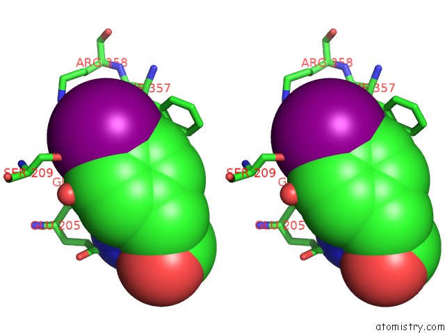
Iodine binding site 2 out of 4 in 1orw
Go back to
Iodine binding site 2 out
of 4 in the Crystal Structure of Porcine Dipeptidyl Peptidase IV (CD26) in Complex with A Peptidomimetic Inhibitor
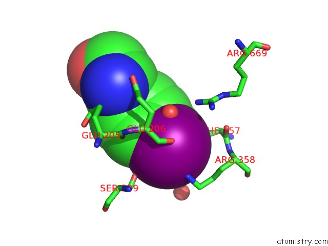
Mono view
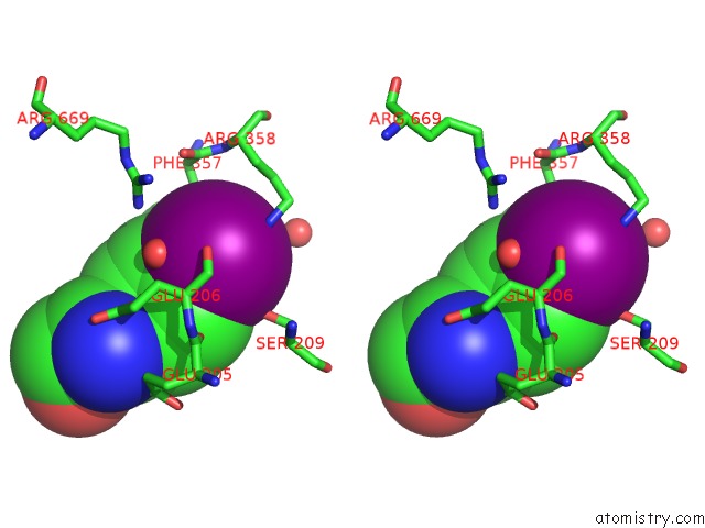
Stereo pair view
Mono view
Stereo pair view
A full contact list of Iodine with other atoms in the I binding
site number 2 of Crystal Structure of Porcine Dipeptidyl Peptidase IV (CD26) in Complex with A Peptidomimetic Inhibitor within 5.0Å range:
|
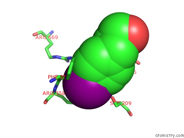
Iodine binding site 3 out of 4 in 1orw
Go back to
Iodine binding site 3 out
of 4 in the Crystal Structure of Porcine Dipeptidyl Peptidase IV (CD26) in Complex with A Peptidomimetic Inhibitor
Mono view
Stereo pair view
Mono view
Stereo pair view
A full contact list of Iodine with other atoms in the I binding
site number 3 of Crystal Structure of Porcine Dipeptidyl Peptidase IV (CD26) in Complex with A Peptidomimetic Inhibitor within 5.0Å range:
|
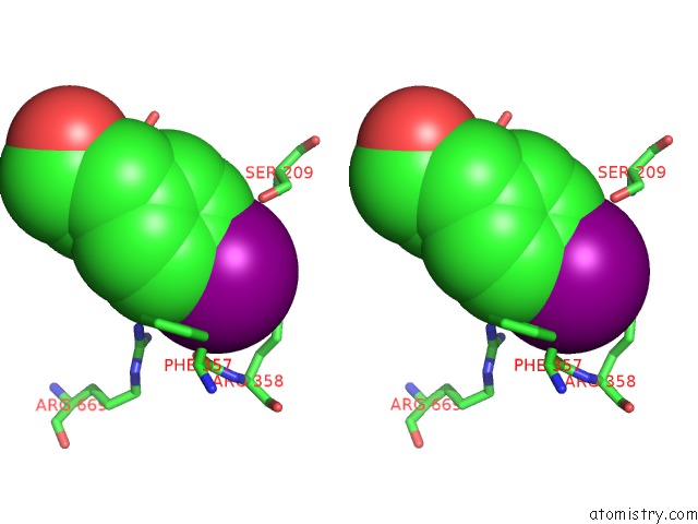
Iodine binding site 4 out of 4 in 1orw
Go back to
Iodine binding site 4 out
of 4 in the Crystal Structure of Porcine Dipeptidyl Peptidase IV (CD26) in Complex with A Peptidomimetic Inhibitor
Mono view
Stereo pair view
Mono view
Stereo pair view
A full contact list of Iodine with other atoms in the I binding
site number 4 of Crystal Structure of Porcine Dipeptidyl Peptidase IV (CD26) in Complex with A Peptidomimetic Inhibitor within 5.0Å range:
|
Reference:
M.Engel,
T.Hoffmann,
L.Wagner,
M.Wermann,
U.Heiser,
R.Kiefersauer,
R.Huber,
W.Bode,
H.U.Demuth,
H.Brandstetter.
The Crystal Structure of Dipeptidyl Peptidase IV (CD26) Reveals Its Functional Regulation and Enzymatic Mechanism Proc.Natl.Acad.Sci.Usa V. 100 5063 2003.
ISSN: ISSN 0027-8424
PubMed: 12690074
DOI: 10.1073/PNAS.0230620100
Page generated: Fri Aug 8 12:07:13 2025
ISSN: ISSN 0027-8424
PubMed: 12690074
DOI: 10.1073/PNAS.0230620100
Last articles
I in 5EIJI in 5EI1
I in 5DLV
I in 5EHB
I in 5E5Y
I in 5E5T
I in 5DN5
I in 5DO6
I in 5DN4
I in 5DLW