Iodine »
PDB 2axe-2fwh »
2d97 »
Iodine in PDB 2d97: Structure of Vil-Xylanase
Enzymatic activity of Structure of Vil-Xylanase
All present enzymatic activity of Structure of Vil-Xylanase:
3.2.1.8;
3.2.1.8;
Protein crystallography data
The structure of Structure of Vil-Xylanase, PDB code: 2d97
was solved by
H.Miyatake,
T.Hasegawa,
A.Yamano,
with X-Ray Crystallography technique. A brief refinement statistics is given in the table below:
| Resolution Low / High (Å) | 18.98 / 2.01 |
| Space group | P 1 21 1 |
| Cell size a, b, c (Å), α, β, γ (°) | 40.347, 38.585, 57.162, 90.00, 110.31, 90.00 |
| R / Rfree (%) | 19.8 / 22.8 |
Iodine Binding Sites:
The binding sites of Iodine atom in the Structure of Vil-Xylanase
(pdb code 2d97). This binding sites where shown within
5.0 Angstroms radius around Iodine atom.
In total 5 binding sites of Iodine where determined in the Structure of Vil-Xylanase, PDB code: 2d97:
Jump to Iodine binding site number: 1; 2; 3; 4; 5;
In total 5 binding sites of Iodine where determined in the Structure of Vil-Xylanase, PDB code: 2d97:
Jump to Iodine binding site number: 1; 2; 3; 4; 5;
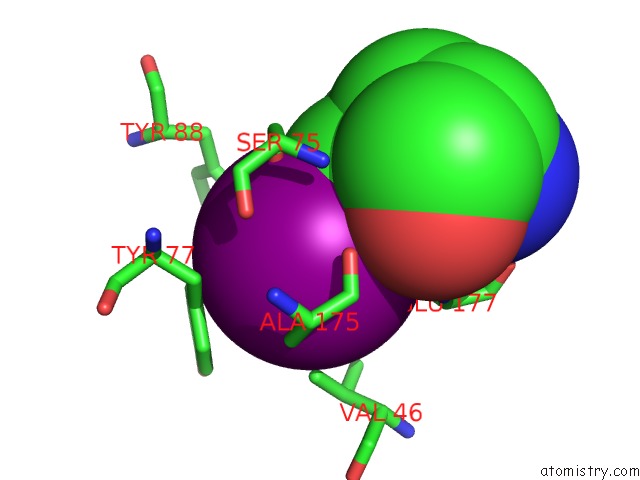
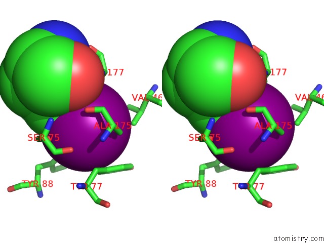
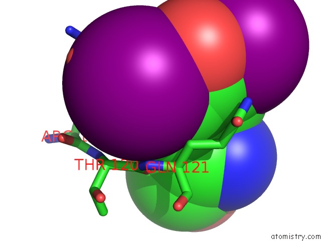
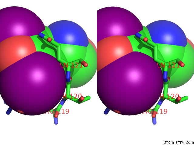
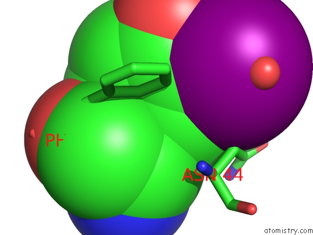
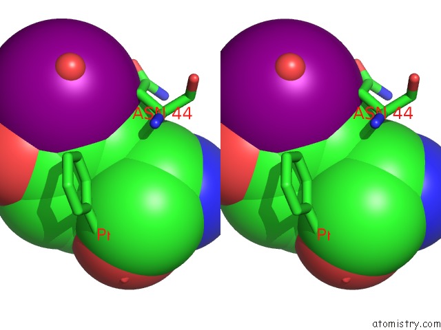
Iodine binding site 1 out of 5 in 2d97
Go back to
Iodine binding site 1 out
of 5 in the Structure of Vil-Xylanase
Mono view
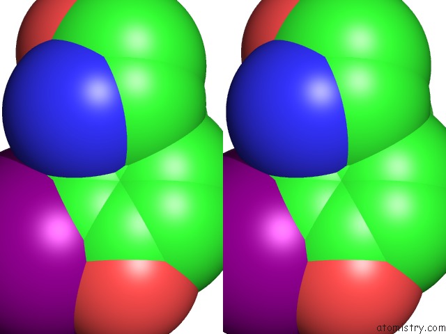
Stereo pair view
Mono view
Stereo pair view
A full contact list of Iodine with other atoms in the I binding
site number 1 of Structure of Vil-Xylanase within 5.0Å range:
|
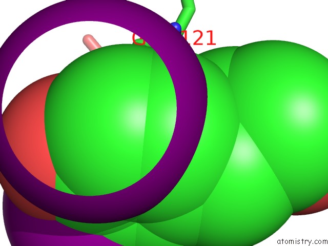
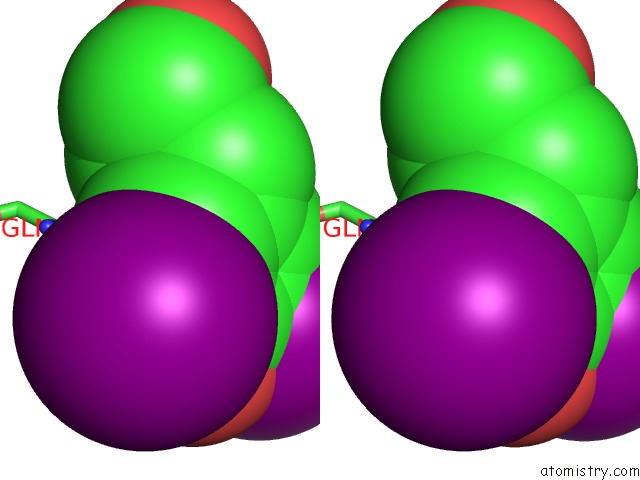
Iodine binding site 2 out of 5 in 2d97
Go back to
Iodine binding site 2 out
of 5 in the Structure of Vil-Xylanase
Mono view
Stereo pair view
Mono view
Stereo pair view
A full contact list of Iodine with other atoms in the I binding
site number 2 of Structure of Vil-Xylanase within 5.0Å range:
|
Iodine binding site 3 out of 5 in 2d97
Go back to
Iodine binding site 3 out
of 5 in the Structure of Vil-Xylanase
Mono view
Stereo pair view
Mono view
Stereo pair view
A full contact list of Iodine with other atoms in the I binding
site number 3 of Structure of Vil-Xylanase within 5.0Å range:
|
Iodine binding site 4 out of 5 in 2d97
Go back to
Iodine binding site 4 out
of 5 in the Structure of Vil-Xylanase
Mono view
Stereo pair view
Mono view
Stereo pair view
A full contact list of Iodine with other atoms in the I binding
site number 4 of Structure of Vil-Xylanase within 5.0Å range:
|
Iodine binding site 5 out of 5 in 2d97
Go back to
Iodine binding site 5 out
of 5 in the Structure of Vil-Xylanase
Mono view
Stereo pair view
Mono view
Stereo pair view
A full contact list of Iodine with other atoms in the I binding
site number 5 of Structure of Vil-Xylanase within 5.0Å range:
|
Reference:
H.Miyatake,
T.Hasegawa,
A.Yamano.
New Methods to Prepare Iodinated Derivatives By Vaporizing Iodine Labelling (Vil) and Hydrogen Peroxide Vil (Hyper-Vil) Acta Crystallogr.,Sect.D V. 62 280 2006.
ISSN: ISSN 0907-4449
PubMed: 16510975
DOI: 10.1107/S0907444905041909
Page generated: Fri Aug 8 13:03:43 2025
ISSN: ISSN 0907-4449
PubMed: 16510975
DOI: 10.1107/S0907444905041909
Last articles
K in 5WIEK in 5WNN
K in 5WK9
K in 5WK7
K in 5WJN
K in 5WGM
K in 5WJM
K in 5WJ8
K in 5WJ1
K in 5WDL