Iodine »
PDB 2qqt-2y5e »
2r1q »
Iodine in PDB 2r1q: Crystal Structure of Iodinated Human Saposin D in Space Group C2221
Protein crystallography data
The structure of Crystal Structure of Iodinated Human Saposin D in Space Group C2221, PDB code: 2r1q
was solved by
T.Maier,
M.Rossman,
W.Saenger,
with X-Ray Crystallography technique. A brief refinement statistics is given in the table below:
| Resolution Low / High (Å) | 19.35 / 2.50 |
| Space group | C 2 2 21 |
| Cell size a, b, c (Å), α, β, γ (°) | 40.465, 74.879, 66.791, 90.00, 90.00, 90.00 |
| R / Rfree (%) | 24.8 / 27.8 |
Iodine Binding Sites:
The binding sites of Iodine atom in the Crystal Structure of Iodinated Human Saposin D in Space Group C2221
(pdb code 2r1q). This binding sites where shown within
5.0 Angstroms radius around Iodine atom.
In total only one binding site of Iodine was determined in the Crystal Structure of Iodinated Human Saposin D in Space Group C2221, PDB code: 2r1q:
In total only one binding site of Iodine was determined in the Crystal Structure of Iodinated Human Saposin D in Space Group C2221, PDB code: 2r1q:
Iodine binding site 1 out of 1 in 2r1q
Go back to
Iodine binding site 1 out
of 1 in the Crystal Structure of Iodinated Human Saposin D in Space Group C2221
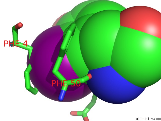
Mono view
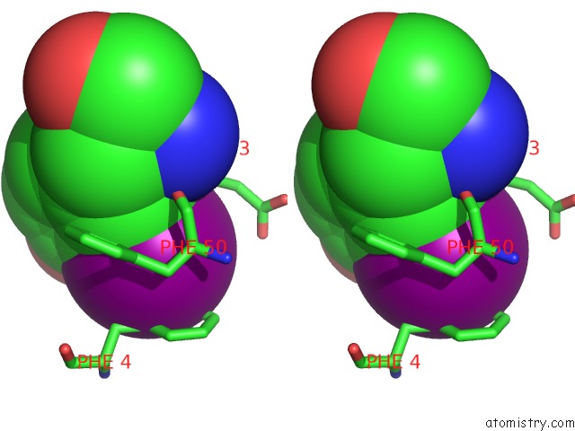
Stereo pair view
Mono view
Stereo pair view
A full contact list of Iodine with other atoms in the I binding
site number 1 of Crystal Structure of Iodinated Human Saposin D in Space Group C2221 within 5.0Å range:
|
Reference:
M.Rossmann,
R.Schultz-Heienbrok,
J.Behlke,
N.Remmel,
C.Alings,
K.Sandhoff,
W.Saenger,
T.Maier.
Crystal Structures of Human Saposins C and D: Implications For Lipid Recognition and Membrane Interactions. Structure V. 16 809 2008.
ISSN: ISSN 0969-2126
PubMed: 18462685
DOI: 10.1016/J.STR.2008.02.016
Page generated: Fri Aug 8 13:35:05 2025
ISSN: ISSN 0969-2126
PubMed: 18462685
DOI: 10.1016/J.STR.2008.02.016
Last articles
Na in 7MD8Na in 7MD9
Na in 7MD6
Na in 7MD1
Na in 7MD0
Na in 7MCY
Na in 7MCP
Na in 7MCQ
Na in 7MCU
Na in 7MCT