Iodine »
PDB 2qqt-2y5e »
2va7 »
Iodine in PDB 2va7: X-Ray Crystal Structure of Beta Secretase Complexed with Compound 27
Enzymatic activity of X-Ray Crystal Structure of Beta Secretase Complexed with Compound 27
All present enzymatic activity of X-Ray Crystal Structure of Beta Secretase Complexed with Compound 27:
3.4.23.46;
3.4.23.46;
Protein crystallography data
The structure of X-Ray Crystal Structure of Beta Secretase Complexed with Compound 27, PDB code: 2va7
was solved by
P.D.Edwards,
J.S.Albert,
M.Sylvester,
D.Aharony,
D.Andisik,
O.Callaghan,
J.B.Campbell,
R.A.Carr,
G.Chessari,
M.Congreve,
M.Frederickson,
R.H.A.Folmer,
S.Geschwindner,
G.Koether,
K.Kolmodin,
J.Krumrine,
R.C.Mauger,
C.W.Murray,
L.L.Olsson,
S.Patel,
N.Spear,
G.Tian,
with X-Ray Crystallography technique. A brief refinement statistics is given in the table below:
| Resolution Low / High (Å) | 47.46 / 2.2 |
| Space group | P 61 2 2 |
| Cell size a, b, c (Å), α, β, γ (°) | 102.687, 102.687, 168.340, 90.00, 90.00, 120.00 |
| R / Rfree (%) | 22.3 / 28 |
Iodine Binding Sites:
The binding sites of Iodine atom in the X-Ray Crystal Structure of Beta Secretase Complexed with Compound 27
(pdb code 2va7). This binding sites where shown within
5.0 Angstroms radius around Iodine atom.
In total 3 binding sites of Iodine where determined in the X-Ray Crystal Structure of Beta Secretase Complexed with Compound 27, PDB code: 2va7:
Jump to Iodine binding site number: 1; 2; 3;
In total 3 binding sites of Iodine where determined in the X-Ray Crystal Structure of Beta Secretase Complexed with Compound 27, PDB code: 2va7:
Jump to Iodine binding site number: 1; 2; 3;
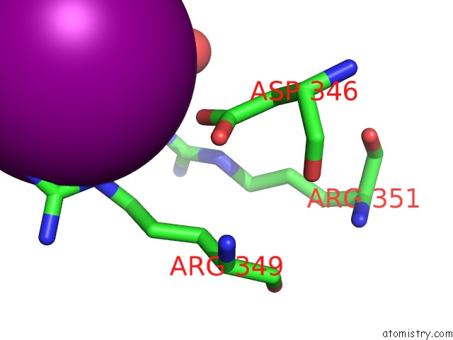
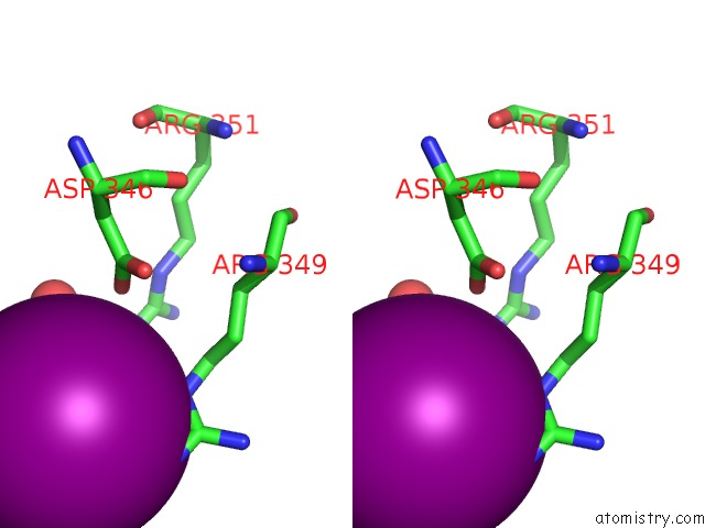
Iodine binding site 1 out of 3 in 2va7
Go back to
Iodine binding site 1 out
of 3 in the X-Ray Crystal Structure of Beta Secretase Complexed with Compound 27
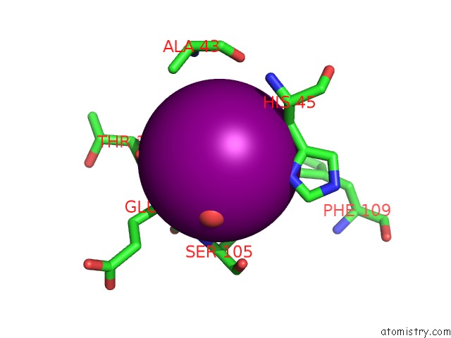
Mono view
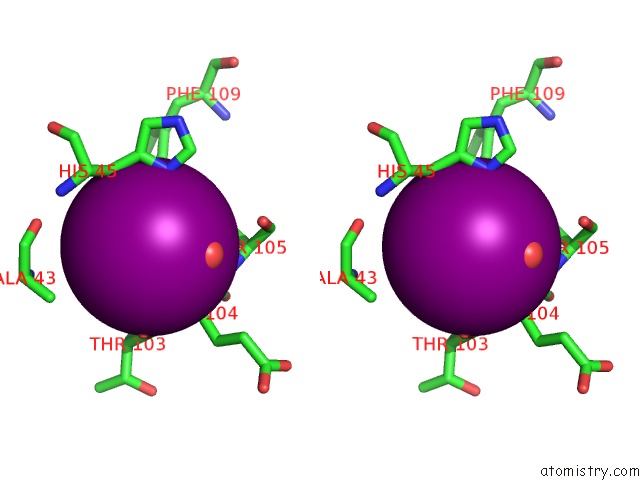
Stereo pair view
Mono view
Stereo pair view
A full contact list of Iodine with other atoms in the I binding
site number 1 of X-Ray Crystal Structure of Beta Secretase Complexed with Compound 27 within 5.0Å range:
|
Iodine binding site 2 out of 3 in 2va7
Go back to
Iodine binding site 2 out
of 3 in the X-Ray Crystal Structure of Beta Secretase Complexed with Compound 27
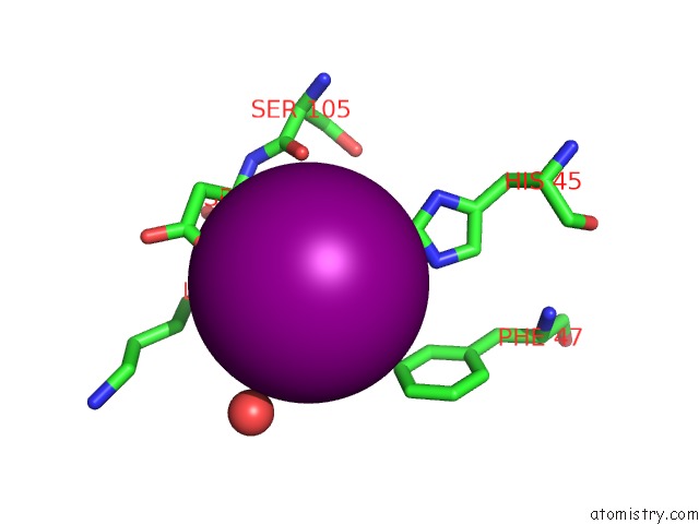
Mono view
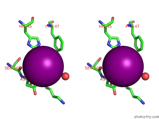
Stereo pair view
Mono view
Stereo pair view
A full contact list of Iodine with other atoms in the I binding
site number 2 of X-Ray Crystal Structure of Beta Secretase Complexed with Compound 27 within 5.0Å range:
|
Iodine binding site 3 out of 3 in 2va7
Go back to
Iodine binding site 3 out
of 3 in the X-Ray Crystal Structure of Beta Secretase Complexed with Compound 27
Mono view
Stereo pair view
Mono view
Stereo pair view
A full contact list of Iodine with other atoms in the I binding
site number 3 of X-Ray Crystal Structure of Beta Secretase Complexed with Compound 27 within 5.0Å range:
|
Reference:
P.D.Edwards,
J.S.Albert,
M.Sylvester,
D.Aharony,
D.Andisik,
O.Callaghan,
J.B.Campbell,
R.A.Carr,
G.Chessari,
M.Congreve,
M.Frederickson,
R.H.A.Folmer,
S.Geschwindner,
G.Koether,
K.Kolmodin,
J.Krumrine,
R.C.Mauger,
C.W.Murray,
L.L.Olsson,
S.Patel,
N.Spear,
G.Tian.
Application of Fragment-Based Lead Generation to the Discovery of Novel, Cyclic Amidine Beta- Secretase Inhibitors with Nanomolar Potency, Cellular Activity, and High Ligand Efficiency. J.Med.Chem. V. 50 5912 2007.
ISSN: ISSN 0022-2623
PubMed: 17985862
DOI: 10.1021/JM070829P
Page generated: Fri Aug 8 13:37:45 2025
ISSN: ISSN 0022-2623
PubMed: 17985862
DOI: 10.1021/JM070829P
Last articles
Na in 3PZBNa in 3PXS
Na in 3PYX
Na in 3PYM
Na in 3PXT
Na in 3PXW
Na in 3PNX
Na in 3PWS
Na in 3PQH
Na in 3PWM