Iodine »
PDB 2y6q-3cgp »
3bas »
Iodine in PDB 3bas: Crystal Structure of the N-Terminal Region of the Scallop Myosin Rod, Monoclinic (C2) Form
Protein crystallography data
The structure of Crystal Structure of the N-Terminal Region of the Scallop Myosin Rod, Monoclinic (C2) Form, PDB code: 3bas
was solved by
J.H.Brown,
C.Cohen,
with X-Ray Crystallography technique. A brief refinement statistics is given in the table below:
| Resolution Low / High (Å) | 20.00 / 2.30 |
| Space group | C 1 2 1 |
| Cell size a, b, c (Å), α, β, γ (°) | 123.335, 44.912, 39.878, 90.00, 95.18, 90.00 |
| R / Rfree (%) | 24.6 / 28.7 |
Iodine Binding Sites:
The binding sites of Iodine atom in the Crystal Structure of the N-Terminal Region of the Scallop Myosin Rod, Monoclinic (C2) Form
(pdb code 3bas). This binding sites where shown within
5.0 Angstroms radius around Iodine atom.
In total only one binding site of Iodine was determined in the Crystal Structure of the N-Terminal Region of the Scallop Myosin Rod, Monoclinic (C2) Form, PDB code: 3bas:
In total only one binding site of Iodine was determined in the Crystal Structure of the N-Terminal Region of the Scallop Myosin Rod, Monoclinic (C2) Form, PDB code: 3bas:
Iodine binding site 1 out of 1 in 3bas
Go back to
Iodine binding site 1 out
of 1 in the Crystal Structure of the N-Terminal Region of the Scallop Myosin Rod, Monoclinic (C2) Form
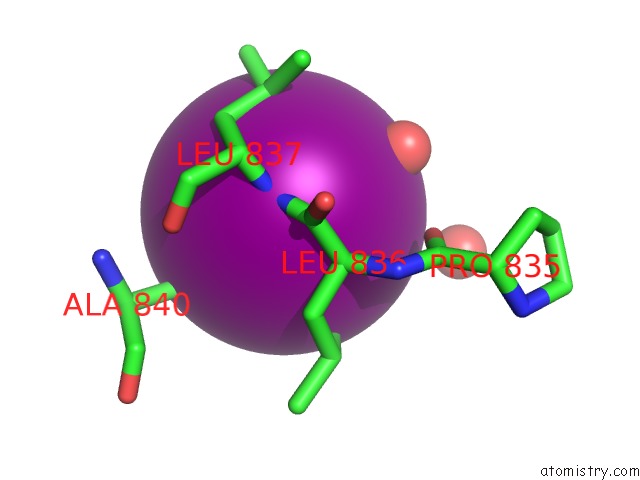
Mono view

Stereo pair view
Mono view
Stereo pair view
A full contact list of Iodine with other atoms in the I binding
site number 1 of Crystal Structure of the N-Terminal Region of the Scallop Myosin Rod, Monoclinic (C2) Form within 5.0Å range:
|
Reference:
J.H.Brown,
Y.Yang,
L.Reshetnikova,
S.Gourinath,
D.Suveges,
J.Kardos,
F.Hobor,
R.Reutzel,
L.Nyitray,
C.Cohen.
An Unstable Head-Rod Junction May Promote Folding Into the Compact Off-State Conformation of Regulated Myosins. J.Mol.Biol. V. 375 1434 2008.
ISSN: ISSN 0022-2836
PubMed: 18155233
DOI: 10.1016/J.JMB.2007.11.071
Page generated: Fri Aug 8 13:58:34 2025
ISSN: ISSN 0022-2836
PubMed: 18155233
DOI: 10.1016/J.JMB.2007.11.071
Last articles
W in 1DV4W in 1FR3
W in 1GUG
W in 1H9R
W in 1H9K
W in 1H0H
W in 1FEZ
W in 1FKA
W in 1E3P
W in 1E18