Iodine »
PDB 3cjq-3fnl »
3fgv »
Iodine in PDB 3fgv: Crystal Structure of A Putative Antibiotic Biosynthesis Monooxygenase (SPO2313) From Silicibacter Pomeroyi Dss-3 at 1.30 A Resolution
Protein crystallography data
The structure of Crystal Structure of A Putative Antibiotic Biosynthesis Monooxygenase (SPO2313) From Silicibacter Pomeroyi Dss-3 at 1.30 A Resolution, PDB code: 3fgv
was solved by
Joint Center For Structural Genomics (Jcsg),
with X-Ray Crystallography technique. A brief refinement statistics is given in the table below:
| Resolution Low / High (Å) | 26.12 / 1.30 |
| Space group | P 65 |
| Cell size a, b, c (Å), α, β, γ (°) | 78.760, 78.760, 69.810, 90.00, 90.00, 120.00 |
| R / Rfree (%) | 13 / 15 |
Iodine Binding Sites:
The binding sites of Iodine atom in the Crystal Structure of A Putative Antibiotic Biosynthesis Monooxygenase (SPO2313) From Silicibacter Pomeroyi Dss-3 at 1.30 A Resolution
(pdb code 3fgv). This binding sites where shown within
5.0 Angstroms radius around Iodine atom.
In total 2 binding sites of Iodine where determined in the Crystal Structure of A Putative Antibiotic Biosynthesis Monooxygenase (SPO2313) From Silicibacter Pomeroyi Dss-3 at 1.30 A Resolution, PDB code: 3fgv:
Jump to Iodine binding site number: 1; 2;
In total 2 binding sites of Iodine where determined in the Crystal Structure of A Putative Antibiotic Biosynthesis Monooxygenase (SPO2313) From Silicibacter Pomeroyi Dss-3 at 1.30 A Resolution, PDB code: 3fgv:
Jump to Iodine binding site number: 1; 2;
Iodine binding site 1 out of 2 in 3fgv
Go back to
Iodine binding site 1 out
of 2 in the Crystal Structure of A Putative Antibiotic Biosynthesis Monooxygenase (SPO2313) From Silicibacter Pomeroyi Dss-3 at 1.30 A Resolution
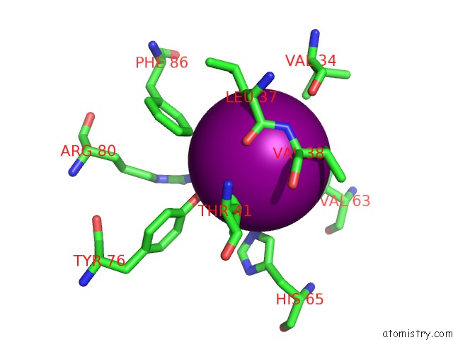
Mono view
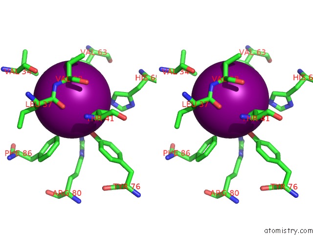
Stereo pair view
Mono view
Stereo pair view
A full contact list of Iodine with other atoms in the I binding
site number 1 of Crystal Structure of A Putative Antibiotic Biosynthesis Monooxygenase (SPO2313) From Silicibacter Pomeroyi Dss-3 at 1.30 A Resolution within 5.0Å range:
|
Iodine binding site 2 out of 2 in 3fgv
Go back to
Iodine binding site 2 out
of 2 in the Crystal Structure of A Putative Antibiotic Biosynthesis Monooxygenase (SPO2313) From Silicibacter Pomeroyi Dss-3 at 1.30 A Resolution
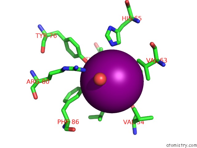
Mono view
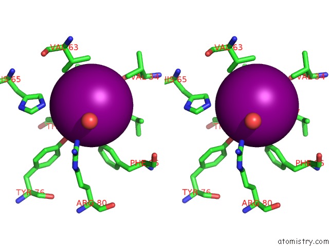
Stereo pair view
Mono view
Stereo pair view
A full contact list of Iodine with other atoms in the I binding
site number 2 of Crystal Structure of A Putative Antibiotic Biosynthesis Monooxygenase (SPO2313) From Silicibacter Pomeroyi Dss-3 at 1.30 A Resolution within 5.0Å range:
|
Reference:
Joint Center For Structural Genomics (Jcsg),
Joint Center For Structural Genomics (Jcsg).
N/A N/A.
Page generated: Sun Aug 11 15:10:42 2024
Last articles
Fe in 7BI7Fe in 7BHC
Fe in 7BIU
Fe in 7BI1
Fe in 7B9A
Fe in 7BGI
Fe in 7BHB
Fe in 7B97
Fe in 7BHA
Fe in 7BBT