Iodine »
PDB 3usl-3zzz »
3w2z »
Iodine in PDB 3w2z: Crystal Structure of the Cyanobacterial Protein
Protein crystallography data
The structure of Crystal Structure of the Cyanobacterial Protein, PDB code: 3w2z
was solved by
R.Narikawa,
N.Muraki,
T.Shiba,
G.Kurisu,
M.Ikeuchi,
with X-Ray Crystallography technique. A brief refinement statistics is given in the table below:
| Resolution Low / High (Å) | 28.31 / 1.80 |
| Space group | P 43 21 2 |
| Cell size a, b, c (Å), α, β, γ (°) | 69.133, 69.133, 124.110, 90.00, 90.00, 90.00 |
| R / Rfree (%) | 19.7 / 22.4 |
Iodine Binding Sites:
The binding sites of Iodine atom in the Crystal Structure of the Cyanobacterial Protein
(pdb code 3w2z). This binding sites where shown within
5.0 Angstroms radius around Iodine atom.
In total only one binding site of Iodine was determined in the Crystal Structure of the Cyanobacterial Protein, PDB code: 3w2z:
In total only one binding site of Iodine was determined in the Crystal Structure of the Cyanobacterial Protein, PDB code: 3w2z:
Iodine binding site 1 out of 1 in 3w2z
Go back to
Iodine binding site 1 out
of 1 in the Crystal Structure of the Cyanobacterial Protein

Mono view
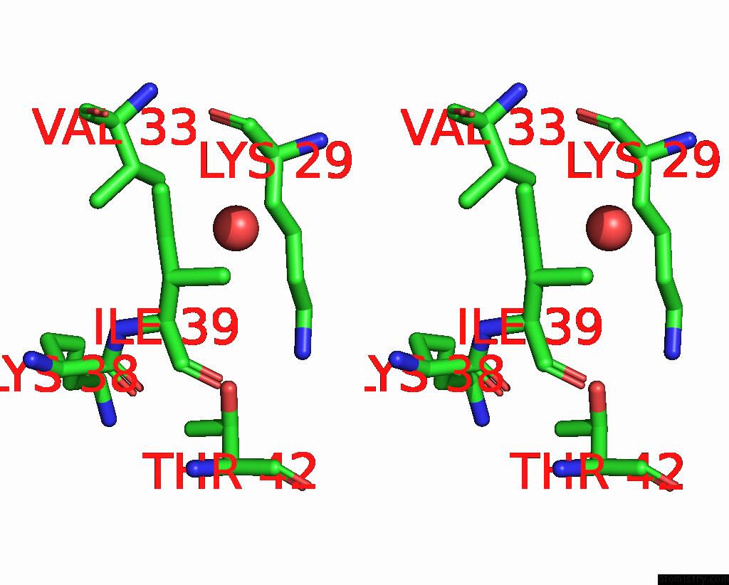
Stereo pair view
Mono view
Stereo pair view
A full contact list of Iodine with other atoms in the I binding
site number 1 of Crystal Structure of the Cyanobacterial Protein within 5.0Å range:
|
Reference:
R.Narikawa,
T.Ishizuka,
N.Muraki,
T.Shiba,
G.Kurisu,
M.Ikeuchi.
Structures of Cyanobacteriochromes From Phototaxis Regulators Anpixj and Tepixj Reveal General and Specific Photoconversion Mechanism Proc.Natl.Acad.Sci.Usa V. 110 918 2013.
ISSN: ISSN 0027-8424
PubMed: 23256156
DOI: 10.1073/PNAS.1212098110
Page generated: Sun Aug 11 17:08:44 2024
ISSN: ISSN 0027-8424
PubMed: 23256156
DOI: 10.1073/PNAS.1212098110
Last articles
Cl in 8RQDCl in 8RRK
Cl in 8RQH
Cl in 8RPM
Cl in 8RPR
Cl in 8RPA
Cl in 8RPB
Cl in 8ROP
Cl in 8RLY
Cl in 8RNX