Iodine »
PDB 4hkl-4jxj »
4j2v »
Iodine in PDB 4j2v: Crystal Structure of Equine Serum Albumin in Complex with 3,5- Diiodosalicylic Acid
Protein crystallography data
The structure of Crystal Structure of Equine Serum Albumin in Complex with 3,5- Diiodosalicylic Acid, PDB code: 4j2v
was solved by
B.Sekula,
A.Bujacz,
K.Zielinski,
G.Bujacz,
with X-Ray Crystallography technique. A brief refinement statistics is given in the table below:
| Resolution Low / High (Å) | 33.39 / 2.12 |
| Space group | P 61 |
| Cell size a, b, c (Å), α, β, γ (°) | 88.880, 88.880, 134.340, 90.00, 90.00, 120.00 |
| R / Rfree (%) | 18.1 / 23.8 |
Iodine Binding Sites:
The binding sites of Iodine atom in the Crystal Structure of Equine Serum Albumin in Complex with 3,5- Diiodosalicylic Acid
(pdb code 4j2v). This binding sites where shown within
5.0 Angstroms radius around Iodine atom.
In total 8 binding sites of Iodine where determined in the Crystal Structure of Equine Serum Albumin in Complex with 3,5- Diiodosalicylic Acid, PDB code: 4j2v:
Jump to Iodine binding site number: 1; 2; 3; 4; 5; 6; 7; 8;
In total 8 binding sites of Iodine where determined in the Crystal Structure of Equine Serum Albumin in Complex with 3,5- Diiodosalicylic Acid, PDB code: 4j2v:
Jump to Iodine binding site number: 1; 2; 3; 4; 5; 6; 7; 8;
Iodine binding site 1 out of 8 in 4j2v
Go back to
Iodine binding site 1 out
of 8 in the Crystal Structure of Equine Serum Albumin in Complex with 3,5- Diiodosalicylic Acid

Mono view
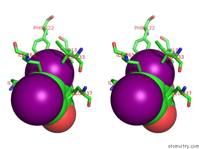
Stereo pair view
Mono view
Stereo pair view
A full contact list of Iodine with other atoms in the I binding
site number 1 of Crystal Structure of Equine Serum Albumin in Complex with 3,5- Diiodosalicylic Acid within 5.0Å range:
|
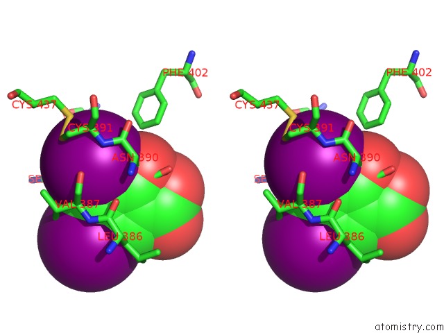
Iodine binding site 2 out of 8 in 4j2v
Go back to
Iodine binding site 2 out
of 8 in the Crystal Structure of Equine Serum Albumin in Complex with 3,5- Diiodosalicylic Acid

Mono view

Stereo pair view
Mono view
Stereo pair view
A full contact list of Iodine with other atoms in the I binding
site number 2 of Crystal Structure of Equine Serum Albumin in Complex with 3,5- Diiodosalicylic Acid within 5.0Å range:
|
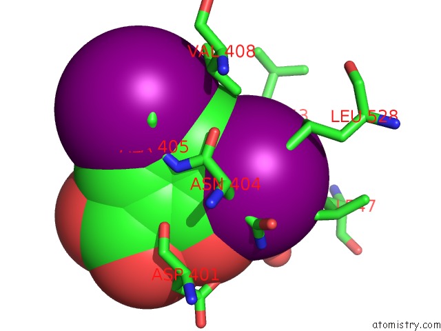
Iodine binding site 3 out of 8 in 4j2v
Go back to
Iodine binding site 3 out
of 8 in the Crystal Structure of Equine Serum Albumin in Complex with 3,5- Diiodosalicylic Acid

Mono view
Stereo pair view
Mono view
Stereo pair view
A full contact list of Iodine with other atoms in the I binding
site number 3 of Crystal Structure of Equine Serum Albumin in Complex with 3,5- Diiodosalicylic Acid within 5.0Å range:
|
Iodine binding site 4 out of 8 in 4j2v
Go back to
Iodine binding site 4 out
of 8 in the Crystal Structure of Equine Serum Albumin in Complex with 3,5- Diiodosalicylic Acid

Mono view

Stereo pair view
Mono view
Stereo pair view
A full contact list of Iodine with other atoms in the I binding
site number 4 of Crystal Structure of Equine Serum Albumin in Complex with 3,5- Diiodosalicylic Acid within 5.0Å range:
|
Iodine binding site 5 out of 8 in 4j2v
Go back to
Iodine binding site 5 out
of 8 in the Crystal Structure of Equine Serum Albumin in Complex with 3,5- Diiodosalicylic Acid

Mono view

Stereo pair view
Mono view
Stereo pair view
A full contact list of Iodine with other atoms in the I binding
site number 5 of Crystal Structure of Equine Serum Albumin in Complex with 3,5- Diiodosalicylic Acid within 5.0Å range:
|
Iodine binding site 6 out of 8 in 4j2v
Go back to
Iodine binding site 6 out
of 8 in the Crystal Structure of Equine Serum Albumin in Complex with 3,5- Diiodosalicylic Acid

Mono view

Stereo pair view
Mono view
Stereo pair view
A full contact list of Iodine with other atoms in the I binding
site number 6 of Crystal Structure of Equine Serum Albumin in Complex with 3,5- Diiodosalicylic Acid within 5.0Å range:
|
Iodine binding site 7 out of 8 in 4j2v
Go back to
Iodine binding site 7 out
of 8 in the Crystal Structure of Equine Serum Albumin in Complex with 3,5- Diiodosalicylic Acid
Mono view

Stereo pair view
Mono view
Stereo pair view
A full contact list of Iodine with other atoms in the I binding
site number 7 of Crystal Structure of Equine Serum Albumin in Complex with 3,5- Diiodosalicylic Acid within 5.0Å range:
|
Iodine binding site 8 out of 8 in 4j2v
Go back to
Iodine binding site 8 out
of 8 in the Crystal Structure of Equine Serum Albumin in Complex with 3,5- Diiodosalicylic Acid

Mono view

Stereo pair view
Mono view
Stereo pair view
A full contact list of Iodine with other atoms in the I binding
site number 8 of Crystal Structure of Equine Serum Albumin in Complex with 3,5- Diiodosalicylic Acid within 5.0Å range:
|
Reference:
B.Sekula,
K.Zielinski,
A.Bujacz.
Crystallographic Studies of the Complexes of Bovine and Equine Serum Albumin with 3,5-Diiodosalicylic Acid. Int.J.Biol.Macromol. V. 60C 316 2013.
ISSN: ISSN 0141-8130
PubMed: 23769932
DOI: 10.1016/J.IJBIOMAC.2013.06.004
Page generated: Fri Aug 8 17:27:12 2025
ISSN: ISSN 0141-8130
PubMed: 23769932
DOI: 10.1016/J.IJBIOMAC.2013.06.004
Last articles
K in 2HJFK in 2HOE
K in 2HJ6
K in 2HIT
K in 2HHK
K in 2H8P
K in 2HH1
K in 2HG9
K in 2HG3
K in 2HDU