Iodine »
PDB 4n6d-4p4z »
4na4 »
Iodine in PDB 4na4: Crystal Structure of Mouse Poly(Adp-Ribose) Glycohydrolase (Parg) Catalytic Domain with Adp-Hpd
Enzymatic activity of Crystal Structure of Mouse Poly(Adp-Ribose) Glycohydrolase (Parg) Catalytic Domain with Adp-Hpd
All present enzymatic activity of Crystal Structure of Mouse Poly(Adp-Ribose) Glycohydrolase (Parg) Catalytic Domain with Adp-Hpd:
3.2.1.143;
3.2.1.143;
Protein crystallography data
The structure of Crystal Structure of Mouse Poly(Adp-Ribose) Glycohydrolase (Parg) Catalytic Domain with Adp-Hpd, PDB code: 4na4
was solved by
Z.Wang,
Z.Cheng,
W.Xu,
with X-Ray Crystallography technique. A brief refinement statistics is given in the table below:
| Resolution Low / High (Å) | 40.00 / 2.50 |
| Space group | P 21 21 2 |
| Cell size a, b, c (Å), α, β, γ (°) | 188.949, 55.569, 165.993, 90.00, 90.00, 90.00 |
| R / Rfree (%) | 24.2 / 28.2 |
Iodine Binding Sites:
The binding sites of Iodine atom in the Crystal Structure of Mouse Poly(Adp-Ribose) Glycohydrolase (Parg) Catalytic Domain with Adp-Hpd
(pdb code 4na4). This binding sites where shown within
5.0 Angstroms radius around Iodine atom.
In total 5 binding sites of Iodine where determined in the Crystal Structure of Mouse Poly(Adp-Ribose) Glycohydrolase (Parg) Catalytic Domain with Adp-Hpd, PDB code: 4na4:
Jump to Iodine binding site number: 1; 2; 3; 4; 5;
In total 5 binding sites of Iodine where determined in the Crystal Structure of Mouse Poly(Adp-Ribose) Glycohydrolase (Parg) Catalytic Domain with Adp-Hpd, PDB code: 4na4:
Jump to Iodine binding site number: 1; 2; 3; 4; 5;
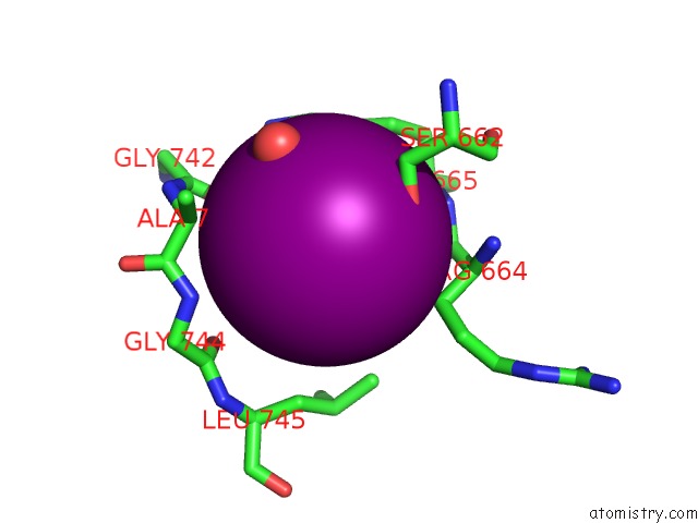
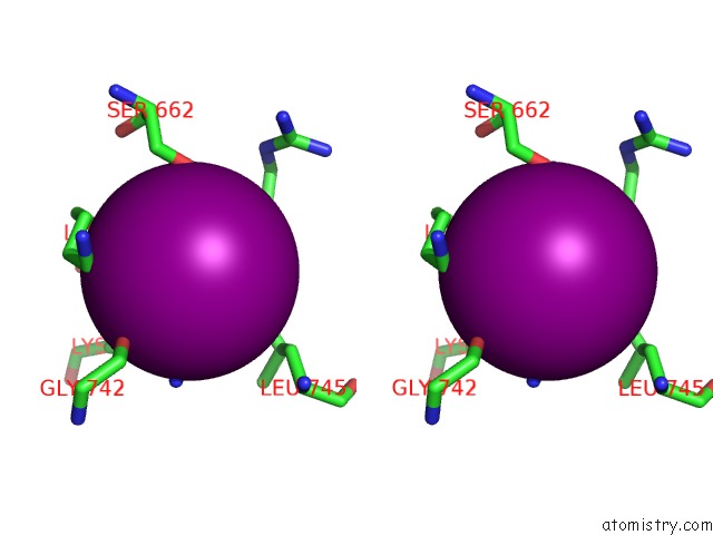
Iodine binding site 1 out of 5 in 4na4
Go back to
Iodine binding site 1 out
of 5 in the Crystal Structure of Mouse Poly(Adp-Ribose) Glycohydrolase (Parg) Catalytic Domain with Adp-Hpd

Mono view

Stereo pair view
Mono view
Stereo pair view
A full contact list of Iodine with other atoms in the I binding
site number 1 of Crystal Structure of Mouse Poly(Adp-Ribose) Glycohydrolase (Parg) Catalytic Domain with Adp-Hpd within 5.0Å range:
|
Iodine binding site 2 out of 5 in 4na4
Go back to
Iodine binding site 2 out
of 5 in the Crystal Structure of Mouse Poly(Adp-Ribose) Glycohydrolase (Parg) Catalytic Domain with Adp-Hpd
Mono view

Stereo pair view
Mono view
Stereo pair view
A full contact list of Iodine with other atoms in the I binding
site number 2 of Crystal Structure of Mouse Poly(Adp-Ribose) Glycohydrolase (Parg) Catalytic Domain with Adp-Hpd within 5.0Å range:
|
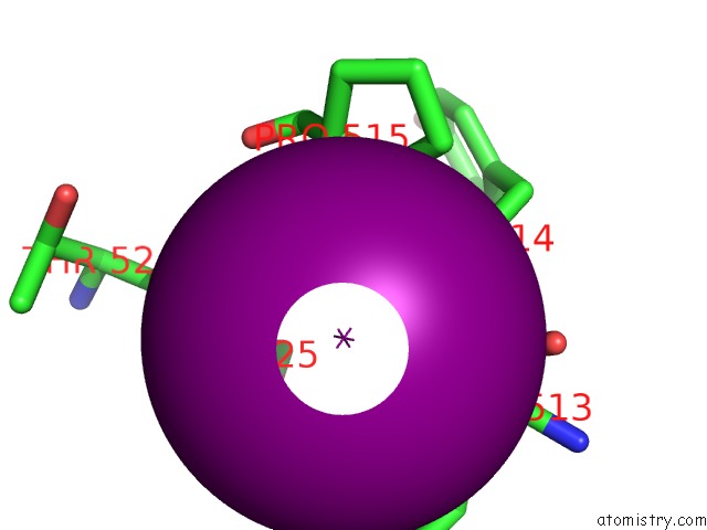
Iodine binding site 3 out of 5 in 4na4
Go back to
Iodine binding site 3 out
of 5 in the Crystal Structure of Mouse Poly(Adp-Ribose) Glycohydrolase (Parg) Catalytic Domain with Adp-Hpd

Mono view
Stereo pair view
Mono view
Stereo pair view
A full contact list of Iodine with other atoms in the I binding
site number 3 of Crystal Structure of Mouse Poly(Adp-Ribose) Glycohydrolase (Parg) Catalytic Domain with Adp-Hpd within 5.0Å range:
|
Iodine binding site 4 out of 5 in 4na4
Go back to
Iodine binding site 4 out
of 5 in the Crystal Structure of Mouse Poly(Adp-Ribose) Glycohydrolase (Parg) Catalytic Domain with Adp-Hpd

Mono view

Stereo pair view
Mono view
Stereo pair view
A full contact list of Iodine with other atoms in the I binding
site number 4 of Crystal Structure of Mouse Poly(Adp-Ribose) Glycohydrolase (Parg) Catalytic Domain with Adp-Hpd within 5.0Å range:
|
Iodine binding site 5 out of 5 in 4na4
Go back to
Iodine binding site 5 out
of 5 in the Crystal Structure of Mouse Poly(Adp-Ribose) Glycohydrolase (Parg) Catalytic Domain with Adp-Hpd
Mono view

Stereo pair view
Mono view
Stereo pair view
A full contact list of Iodine with other atoms in the I binding
site number 5 of Crystal Structure of Mouse Poly(Adp-Ribose) Glycohydrolase (Parg) Catalytic Domain with Adp-Hpd within 5.0Å range:
|
Reference:
Z.Wang,
J.P.Gagne,
G.G.Poirier,
W.Xu.
Crystallographic and Biochemical Analysis of the Mouse Poly(Adp-Ribose) Glycohydrolase. Plos One V. 9 86010 2014.
ISSN: ESSN 1932-6203
PubMed: 24465839
DOI: 10.1371/JOURNAL.PONE.0086010
Page generated: Sun Aug 11 18:53:24 2024
ISSN: ESSN 1932-6203
PubMed: 24465839
DOI: 10.1371/JOURNAL.PONE.0086010
Last articles
Fe in 9KDGFe in 9KDF
Fe in 9K3B
Fe in 9K3Q
Fe in 9K39
Fe in 9K38
Fe in 9JP5
Fe in 9K2G
Fe in 9K36
Fe in 9JN8