Iodine »
PDB 4p9t-4ttc »
4q8d »
Iodine in PDB 4q8d: Crystal Structure of A Macrocyclic Beta-Sheet Peptide Containing Two Beta-Strands From Amyloid Beta Residues 15-23
Protein crystallography data
The structure of Crystal Structure of A Macrocyclic Beta-Sheet Peptide Containing Two Beta-Strands From Amyloid Beta Residues 15-23, PDB code: 4q8d
was solved by
J.D.Pham,
R.K.Spencer,
K.H.Chen,
J.S.Nowick,
with X-Ray Crystallography technique. A brief refinement statistics is given in the table below:
| Resolution Low / High (Å) | 17.56 / 1.75 |
| Space group | C 1 2 1 |
| Cell size a, b, c (Å), α, β, γ (°) | 32.174, 62.852, 20.094, 90.00, 89.98, 90.00 |
| R / Rfree (%) | 17.9 / 22 |
Iodine Binding Sites:
The binding sites of Iodine atom in the Crystal Structure of A Macrocyclic Beta-Sheet Peptide Containing Two Beta-Strands From Amyloid Beta Residues 15-23
(pdb code 4q8d). This binding sites where shown within
5.0 Angstroms radius around Iodine atom.
In total 2 binding sites of Iodine where determined in the Crystal Structure of A Macrocyclic Beta-Sheet Peptide Containing Two Beta-Strands From Amyloid Beta Residues 15-23, PDB code: 4q8d:
Jump to Iodine binding site number: 1; 2;
In total 2 binding sites of Iodine where determined in the Crystal Structure of A Macrocyclic Beta-Sheet Peptide Containing Two Beta-Strands From Amyloid Beta Residues 15-23, PDB code: 4q8d:
Jump to Iodine binding site number: 1; 2;
Iodine binding site 1 out of 2 in 4q8d
Go back to
Iodine binding site 1 out
of 2 in the Crystal Structure of A Macrocyclic Beta-Sheet Peptide Containing Two Beta-Strands From Amyloid Beta Residues 15-23
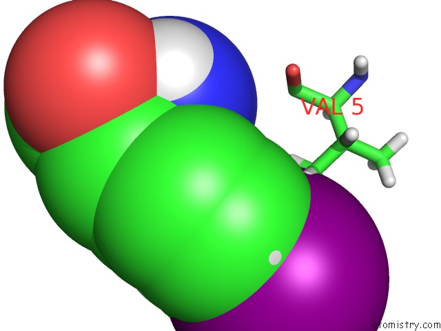
Mono view
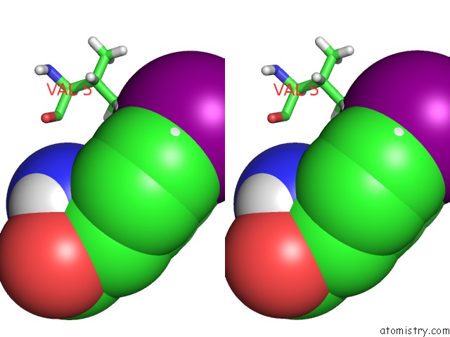
Stereo pair view
Mono view
Stereo pair view
A full contact list of Iodine with other atoms in the I binding
site number 1 of Crystal Structure of A Macrocyclic Beta-Sheet Peptide Containing Two Beta-Strands From Amyloid Beta Residues 15-23 within 5.0Å range:
|
Iodine binding site 2 out of 2 in 4q8d
Go back to
Iodine binding site 2 out
of 2 in the Crystal Structure of A Macrocyclic Beta-Sheet Peptide Containing Two Beta-Strands From Amyloid Beta Residues 15-23
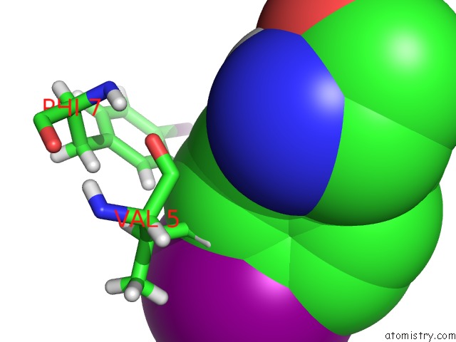
Mono view
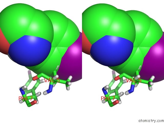
Stereo pair view
Mono view
Stereo pair view
A full contact list of Iodine with other atoms in the I binding
site number 2 of Crystal Structure of A Macrocyclic Beta-Sheet Peptide Containing Two Beta-Strands From Amyloid Beta Residues 15-23 within 5.0Å range:
|
Reference:
J.D.Pham,
R.K.Spencer,
K.H.Chen,
J.S.Nowick.
A Fibril-Like Assembly of Oligomers of A Peptide Derived From Beta-Amyloid. J.Am.Chem.Soc. V. 136 12682 2014.
ISSN: ISSN 0002-7863
PubMed: 25068693
DOI: 10.1021/JA505713Y
Page generated: Fri Aug 8 18:38:01 2025
ISSN: ISSN 0002-7863
PubMed: 25068693
DOI: 10.1021/JA505713Y
Last articles
Na in 8GR6Na in 8GQ0
Na in 8GSG
Na in 8GQZ
Na in 8GPZ
Na in 8GNK
Na in 8GKP
Na in 8GPC
Na in 8GNE
Na in 8GMW