Iodine »
PDB 4p9t-4ttc »
4s2f »
Iodine in PDB 4s2f: Joint X-Ray/Neutron Structure of Trichoderma Reesei Xylanase II at pH 4.4
Enzymatic activity of Joint X-Ray/Neutron Structure of Trichoderma Reesei Xylanase II at pH 4.4
All present enzymatic activity of Joint X-Ray/Neutron Structure of Trichoderma Reesei Xylanase II at pH 4.4:
3.2.1.8;
3.2.1.8;
Protein crystallography data
The structure of Joint X-Ray/Neutron Structure of Trichoderma Reesei Xylanase II at pH 4.4, PDB code: 4s2f
was solved by
A.Kovalevsky,
Q.Wan,
P.Langan,
with X-Ray Crystallography technique. A brief refinement statistics is given in the table below:
| Resolution Low / High (Å) | N/A / 1.70 |
| Space group | P 21 21 21 |
| Cell size a, b, c (Å), α, β, γ (°) | 49.963, 59.621, 70.588, 90.00, 90.00, 90.00 |
| R / Rfree (%) | 26.1 / 30.4 |
Iodine Binding Sites:
The binding sites of Iodine atom in the Joint X-Ray/Neutron Structure of Trichoderma Reesei Xylanase II at pH 4.4
(pdb code 4s2f). This binding sites where shown within
5.0 Angstroms radius around Iodine atom.
In total only one binding site of Iodine was determined in the Joint X-Ray/Neutron Structure of Trichoderma Reesei Xylanase II at pH 4.4, PDB code: 4s2f:
In total only one binding site of Iodine was determined in the Joint X-Ray/Neutron Structure of Trichoderma Reesei Xylanase II at pH 4.4, PDB code: 4s2f:
Iodine binding site 1 out of 1 in 4s2f
Go back to
Iodine binding site 1 out
of 1 in the Joint X-Ray/Neutron Structure of Trichoderma Reesei Xylanase II at pH 4.4
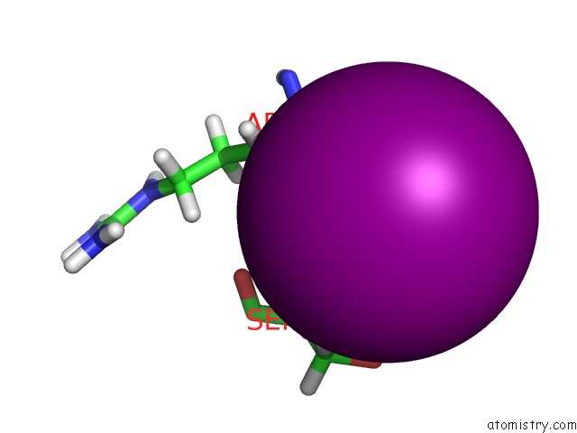
Mono view
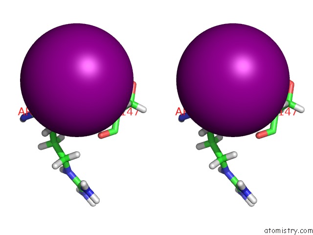
Stereo pair view
Mono view
Stereo pair view
A full contact list of Iodine with other atoms in the I binding
site number 1 of Joint X-Ray/Neutron Structure of Trichoderma Reesei Xylanase II at pH 4.4 within 5.0Å range:
|
Reference:
Q.Wan,
J.M.Parks,
B.L.Hanson,
S.Z.Fisher,
A.Ostermann,
T.E.Schrader,
D.E.Graham,
L.Coates,
P.Langan,
A.Kovalevsky.
Direct Determination of Protonation States and Visualization of Hydrogen Bonding in A Glycoside Hydrolase with Neutron Crystallography. Proc.Natl.Acad.Sci.Usa V. 112 12384 2015.
ISSN: ISSN 0027-8424
PubMed: 26392527
DOI: 10.1073/PNAS.1504986112
Page generated: Fri Aug 8 18:44:07 2025
ISSN: ISSN 0027-8424
PubMed: 26392527
DOI: 10.1073/PNAS.1504986112
Last articles
Na in 8G2ENa in 8G1L
Na in 8FYV
Na in 8G1Q
Na in 8G18
Na in 8G0I
Na in 8FWU
Na in 8FWW
Na in 8FWS
Na in 8FYN