Iodine »
PDB 4xnw-5ax3 »
4xpf »
Iodine in PDB 4xpf: X-Ray Structure of Drosophila Dopamine Transporter with Subsiteb Mutations (D121G/S426M) Bound to Rti-55
Protein crystallography data
The structure of X-Ray Structure of Drosophila Dopamine Transporter with Subsiteb Mutations (D121G/S426M) Bound to Rti-55, PDB code: 4xpf
was solved by
A.Penmatsa,
K.H.Wang,
E.Gouaux,
with X-Ray Crystallography technique. A brief refinement statistics is given in the table below:
| Resolution Low / High (Å) | 48.32 / 3.27 |
| Space group | P 21 21 21 |
| Cell size a, b, c (Å), α, β, γ (°) | 97.415, 140.312, 166.962, 90.00, 90.00, 90.00 |
| R / Rfree (%) | 24.1 / 29.6 |
Other elements in 4xpf:
The structure of X-Ray Structure of Drosophila Dopamine Transporter with Subsiteb Mutations (D121G/S426M) Bound to Rti-55 also contains other interesting chemical elements:
| Chlorine | (Cl) | 1 atom |
| Sodium | (Na) | 2 atoms |
Iodine Binding Sites:
The binding sites of Iodine atom in the X-Ray Structure of Drosophila Dopamine Transporter with Subsiteb Mutations (D121G/S426M) Bound to Rti-55
(pdb code 4xpf). This binding sites where shown within
5.0 Angstroms radius around Iodine atom.
In total only one binding site of Iodine was determined in the X-Ray Structure of Drosophila Dopamine Transporter with Subsiteb Mutations (D121G/S426M) Bound to Rti-55, PDB code: 4xpf:
In total only one binding site of Iodine was determined in the X-Ray Structure of Drosophila Dopamine Transporter with Subsiteb Mutations (D121G/S426M) Bound to Rti-55, PDB code: 4xpf:
Iodine binding site 1 out of 1 in 4xpf
Go back to
Iodine binding site 1 out
of 1 in the X-Ray Structure of Drosophila Dopamine Transporter with Subsiteb Mutations (D121G/S426M) Bound to Rti-55
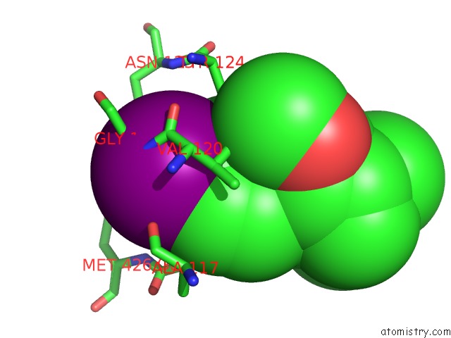
Mono view
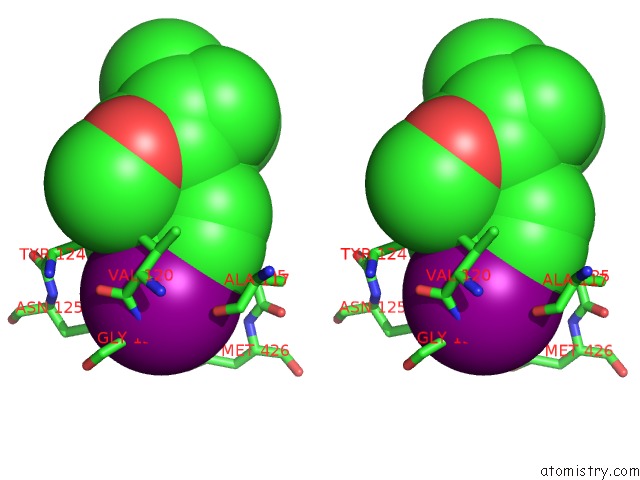
Stereo pair view
Mono view
Stereo pair view
A full contact list of Iodine with other atoms in the I binding
site number 1 of X-Ray Structure of Drosophila Dopamine Transporter with Subsiteb Mutations (D121G/S426M) Bound to Rti-55 within 5.0Å range:
|
Reference:
A.Penmatsa,
K.H.Wang,
E.Gouaux.
Structural Basis For Neurotransmitter and Psycho Stimulant Recognition By the Drosophila Dopamine Transporter Nature 2015.
ISSN: ESSN 1476-4687
Page generated: Fri Aug 8 19:10:04 2025
ISSN: ESSN 1476-4687
Last articles
Na in 1NJ9Na in 1NHX
Na in 1ND4
Na in 1NHJ
Na in 1NAI
Na in 1NAH
Na in 1N82
Na in 1NA0
Na in 1N76
Na in 1N3E