Iodine »
PDB 4xnw-5ax3 »
5aoj »
Iodine in PDB 5aoj: Structure of the P53 Cancer Mutant Y220C in Complex with 2-Hydroxy-3, 5-Diiodo-4-(1H-Pyrrol-1-Yl)Benzoic Acid
Protein crystallography data
The structure of Structure of the P53 Cancer Mutant Y220C in Complex with 2-Hydroxy-3, 5-Diiodo-4-(1H-Pyrrol-1-Yl)Benzoic Acid, PDB code: 5aoj
was solved by
A.C.Joerger,
M.G.Baud,
M.R.Bauer,
A.R.Fersht,
with X-Ray Crystallography technique. A brief refinement statistics is given in the table below:
| Resolution Low / High (Å) | 29.47 / 1.47 |
| Space group | P 21 21 21 |
| Cell size a, b, c (Å), α, β, γ (°) | 65.216, 71.108, 105.353, 90.00, 90.00, 90.00 |
| R / Rfree (%) | 14.9 / 17.8 |
Other elements in 5aoj:
The structure of Structure of the P53 Cancer Mutant Y220C in Complex with 2-Hydroxy-3, 5-Diiodo-4-(1H-Pyrrol-1-Yl)Benzoic Acid also contains other interesting chemical elements:
| Zinc | (Zn) | 2 atoms |
Iodine Binding Sites:
The binding sites of Iodine atom in the Structure of the P53 Cancer Mutant Y220C in Complex with 2-Hydroxy-3, 5-Diiodo-4-(1H-Pyrrol-1-Yl)Benzoic Acid
(pdb code 5aoj). This binding sites where shown within
5.0 Angstroms radius around Iodine atom.
In total 4 binding sites of Iodine where determined in the Structure of the P53 Cancer Mutant Y220C in Complex with 2-Hydroxy-3, 5-Diiodo-4-(1H-Pyrrol-1-Yl)Benzoic Acid, PDB code: 5aoj:
Jump to Iodine binding site number: 1; 2; 3; 4;
In total 4 binding sites of Iodine where determined in the Structure of the P53 Cancer Mutant Y220C in Complex with 2-Hydroxy-3, 5-Diiodo-4-(1H-Pyrrol-1-Yl)Benzoic Acid, PDB code: 5aoj:
Jump to Iodine binding site number: 1; 2; 3; 4;
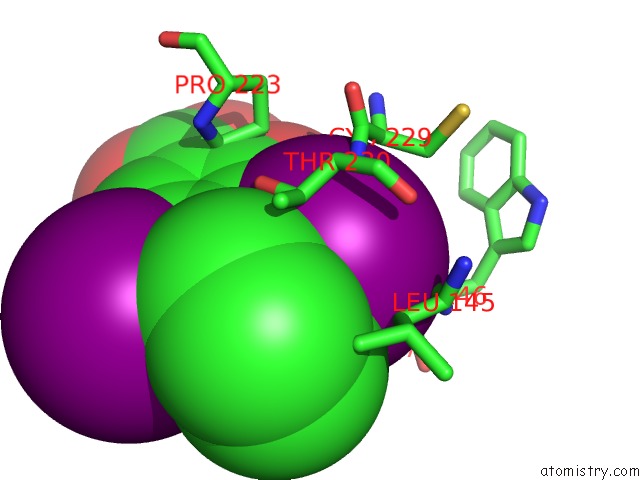
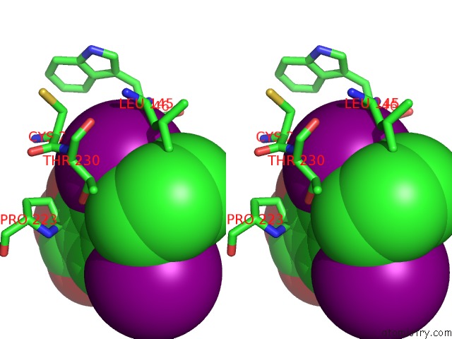
Iodine binding site 1 out of 4 in 5aoj
Go back to
Iodine binding site 1 out
of 4 in the Structure of the P53 Cancer Mutant Y220C in Complex with 2-Hydroxy-3, 5-Diiodo-4-(1H-Pyrrol-1-Yl)Benzoic Acid
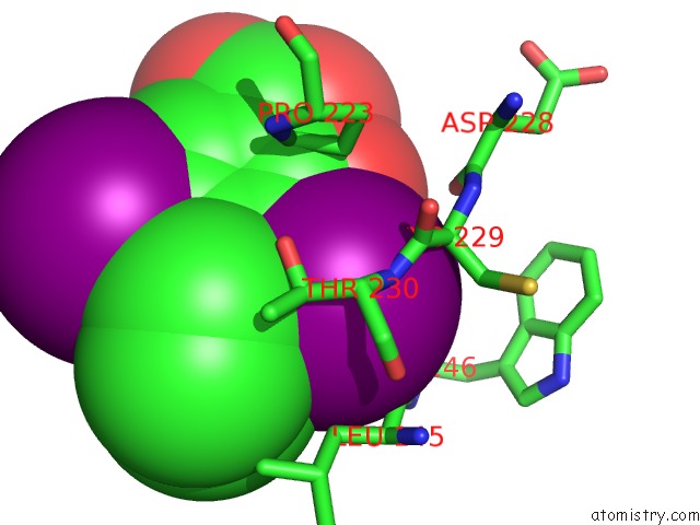
Mono view
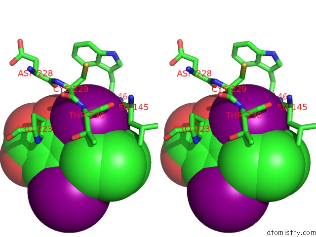
Stereo pair view
Mono view
Stereo pair view
A full contact list of Iodine with other atoms in the I binding
site number 1 of Structure of the P53 Cancer Mutant Y220C in Complex with 2-Hydroxy-3, 5-Diiodo-4-(1H-Pyrrol-1-Yl)Benzoic Acid within 5.0Å range:
|
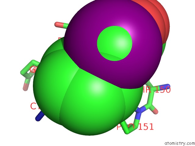
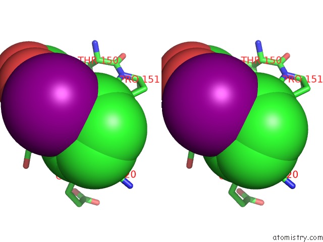
Iodine binding site 2 out of 4 in 5aoj
Go back to
Iodine binding site 2 out
of 4 in the Structure of the P53 Cancer Mutant Y220C in Complex with 2-Hydroxy-3, 5-Diiodo-4-(1H-Pyrrol-1-Yl)Benzoic Acid
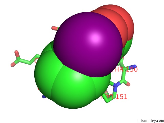
Mono view
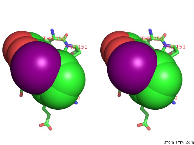
Stereo pair view
Mono view
Stereo pair view
A full contact list of Iodine with other atoms in the I binding
site number 2 of Structure of the P53 Cancer Mutant Y220C in Complex with 2-Hydroxy-3, 5-Diiodo-4-(1H-Pyrrol-1-Yl)Benzoic Acid within 5.0Å range:
|
Iodine binding site 3 out of 4 in 5aoj
Go back to
Iodine binding site 3 out
of 4 in the Structure of the P53 Cancer Mutant Y220C in Complex with 2-Hydroxy-3, 5-Diiodo-4-(1H-Pyrrol-1-Yl)Benzoic Acid
Mono view
Stereo pair view
Mono view
Stereo pair view
A full contact list of Iodine with other atoms in the I binding
site number 3 of Structure of the P53 Cancer Mutant Y220C in Complex with 2-Hydroxy-3, 5-Diiodo-4-(1H-Pyrrol-1-Yl)Benzoic Acid within 5.0Å range:
|
Iodine binding site 4 out of 4 in 5aoj
Go back to
Iodine binding site 4 out
of 4 in the Structure of the P53 Cancer Mutant Y220C in Complex with 2-Hydroxy-3, 5-Diiodo-4-(1H-Pyrrol-1-Yl)Benzoic Acid
Mono view
Stereo pair view
Mono view
Stereo pair view
A full contact list of Iodine with other atoms in the I binding
site number 4 of Structure of the P53 Cancer Mutant Y220C in Complex with 2-Hydroxy-3, 5-Diiodo-4-(1H-Pyrrol-1-Yl)Benzoic Acid within 5.0Å range:
|
Reference:
A.C.Joerger,
M.R.Bauer,
R.Wilcken,
M.G.J.Baud,
H.Harbrecht,
T.E.Exner,
F.M.Boeckler,
J.Spencer,
A.R.Fersht.
Exploiting Transient Protein States For the Design of Small-Molecule Stabilizers of Mutant P53. Structure V. 23 2246 2015.
ISSN: ISSN 0969-2126
PubMed: 26636255
DOI: 10.1016/J.STR.2015.10.016
Page generated: Fri Aug 8 19:19:47 2025
ISSN: ISSN 0969-2126
PubMed: 26636255
DOI: 10.1016/J.STR.2015.10.016
Last articles
Ir in 8OJXIr in 8EXB
Ir in 8EWS
Ir in 8EWR
Ir in 8EWQ
Ir in 8EWP
Ir in 8EWN
Ir in 8EWM
Ir in 7UOR
Ir in 8EWE