Iodine »
PDB 5enk-5kio »
5ijw »
Iodine in PDB 5ijw: Glutamate Racemase (Muri) From Mycobacterium Smegmatis with Bound D- Glutamate, 1.8 Angstrom Resolution, X-Ray Diffraction
Enzymatic activity of Glutamate Racemase (Muri) From Mycobacterium Smegmatis with Bound D- Glutamate, 1.8 Angstrom Resolution, X-Ray Diffraction
All present enzymatic activity of Glutamate Racemase (Muri) From Mycobacterium Smegmatis with Bound D- Glutamate, 1.8 Angstrom Resolution, X-Ray Diffraction:
5.1.1.3;
5.1.1.3;
Protein crystallography data
The structure of Glutamate Racemase (Muri) From Mycobacterium Smegmatis with Bound D- Glutamate, 1.8 Angstrom Resolution, X-Ray Diffraction, PDB code: 5ijw
was solved by
S.Poen,
Y.Nakatani,
K.Krause,
with X-Ray Crystallography technique. A brief refinement statistics is given in the table below:
| Resolution Low / High (Å) | 52.40 / 1.76 |
| Space group | P 21 21 21 |
| Cell size a, b, c (Å), α, β, γ (°) | 61.100, 90.100, 101.700, 90.00, 90.00, 90.00 |
| R / Rfree (%) | 17 / 20.5 |
Iodine Binding Sites:
The binding sites of Iodine atom in the Glutamate Racemase (Muri) From Mycobacterium Smegmatis with Bound D- Glutamate, 1.8 Angstrom Resolution, X-Ray Diffraction
(pdb code 5ijw). This binding sites where shown within
5.0 Angstroms radius around Iodine atom.
In total 3 binding sites of Iodine where determined in the Glutamate Racemase (Muri) From Mycobacterium Smegmatis with Bound D- Glutamate, 1.8 Angstrom Resolution, X-Ray Diffraction, PDB code: 5ijw:
Jump to Iodine binding site number: 1; 2; 3;
In total 3 binding sites of Iodine where determined in the Glutamate Racemase (Muri) From Mycobacterium Smegmatis with Bound D- Glutamate, 1.8 Angstrom Resolution, X-Ray Diffraction, PDB code: 5ijw:
Jump to Iodine binding site number: 1; 2; 3;
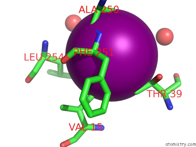
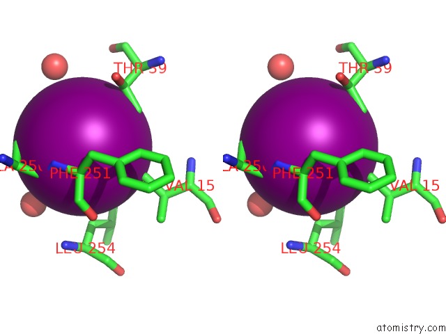
Iodine binding site 1 out of 3 in 5ijw
Go back to
Iodine binding site 1 out
of 3 in the Glutamate Racemase (Muri) From Mycobacterium Smegmatis with Bound D- Glutamate, 1.8 Angstrom Resolution, X-Ray Diffraction
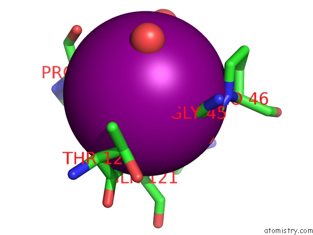
Mono view
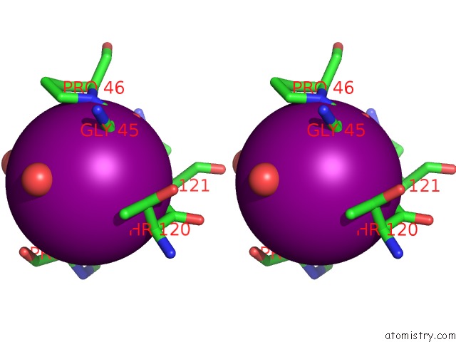
Stereo pair view
Mono view
Stereo pair view
A full contact list of Iodine with other atoms in the I binding
site number 1 of Glutamate Racemase (Muri) From Mycobacterium Smegmatis with Bound D- Glutamate, 1.8 Angstrom Resolution, X-Ray Diffraction within 5.0Å range:
|
Iodine binding site 2 out of 3 in 5ijw
Go back to
Iodine binding site 2 out
of 3 in the Glutamate Racemase (Muri) From Mycobacterium Smegmatis with Bound D- Glutamate, 1.8 Angstrom Resolution, X-Ray Diffraction
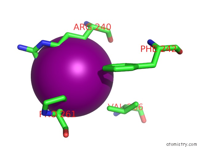
Mono view
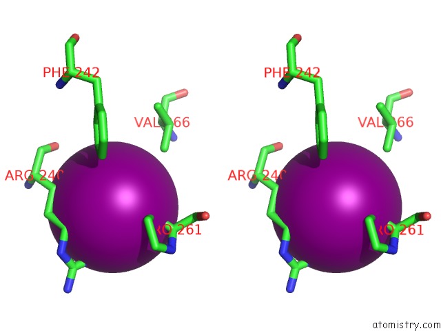
Stereo pair view
Mono view
Stereo pair view
A full contact list of Iodine with other atoms in the I binding
site number 2 of Glutamate Racemase (Muri) From Mycobacterium Smegmatis with Bound D- Glutamate, 1.8 Angstrom Resolution, X-Ray Diffraction within 5.0Å range:
|
Iodine binding site 3 out of 3 in 5ijw
Go back to
Iodine binding site 3 out
of 3 in the Glutamate Racemase (Muri) From Mycobacterium Smegmatis with Bound D- Glutamate, 1.8 Angstrom Resolution, X-Ray Diffraction
Mono view
Stereo pair view
Mono view
Stereo pair view
A full contact list of Iodine with other atoms in the I binding
site number 3 of Glutamate Racemase (Muri) From Mycobacterium Smegmatis with Bound D- Glutamate, 1.8 Angstrom Resolution, X-Ray Diffraction within 5.0Å range:
|
Reference:
S.Poen,
Y.Nakatani,
H.K.Opel-Reading,
M.Lasse,
R.C.Dobson,
K.L.Krause.
Exploring the Structure of Glutamate Racemase From Mycobacterium Tuberculosis As A Template For Anti-Mycobacterial Drug Discovery. Biochem. J. V. 473 1267 2016.
ISSN: ESSN 1470-8728
PubMed: 26964898
DOI: 10.1042/BCJ20160186
Page generated: Sun Aug 11 21:04:50 2024
ISSN: ESSN 1470-8728
PubMed: 26964898
DOI: 10.1042/BCJ20160186
Last articles
I in 5F1WI in 5EOJ
I in 5EUZ
I in 5ENM
I in 5ENK
I in 5EEK
I in 5E9O
I in 5EMS
I in 5EIJ
I in 5EI1