Iodine »
PDB 5sb0-5w1i »
5td5 »
Iodine in PDB 5td5: Crystal Structure of Human APOBEC3B Variant Complexed with Ssdna
Protein crystallography data
The structure of Crystal Structure of Human APOBEC3B Variant Complexed with Ssdna, PDB code: 5td5
was solved by
K.Shi,
S.Banerjee,
K.Kurahashi,
H.Aihara,
with X-Ray Crystallography technique. A brief refinement statistics is given in the table below:
| Resolution Low / High (Å) | 83.49 / 1.72 |
| Space group | P 64 2 2 |
| Cell size a, b, c (Å), α, β, γ (°) | 96.411, 96.411, 84.875, 90.00, 90.00, 120.00 |
| R / Rfree (%) | 18 / 20.9 |
Other elements in 5td5:
The structure of Crystal Structure of Human APOBEC3B Variant Complexed with Ssdna also contains other interesting chemical elements:
| Zinc | (Zn) | 1 atom |
| Chlorine | (Cl) | 2 atoms |
Iodine Binding Sites:
The binding sites of Iodine atom in the Crystal Structure of Human APOBEC3B Variant Complexed with Ssdna
(pdb code 5td5). This binding sites where shown within
5.0 Angstroms radius around Iodine atom.
In total 9 binding sites of Iodine where determined in the Crystal Structure of Human APOBEC3B Variant Complexed with Ssdna, PDB code: 5td5:
Jump to Iodine binding site number: 1; 2; 3; 4; 5; 6; 7; 8; 9;
In total 9 binding sites of Iodine where determined in the Crystal Structure of Human APOBEC3B Variant Complexed with Ssdna, PDB code: 5td5:
Jump to Iodine binding site number: 1; 2; 3; 4; 5; 6; 7; 8; 9;
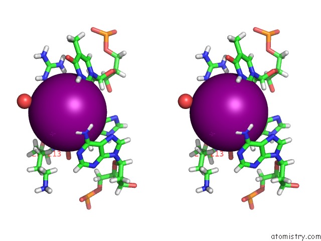
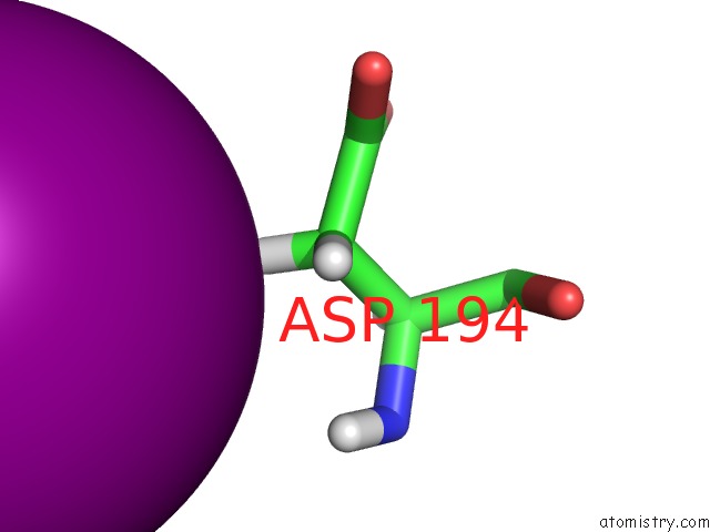
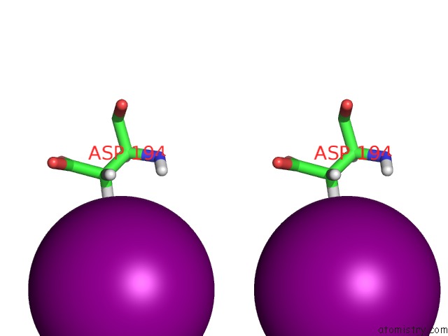
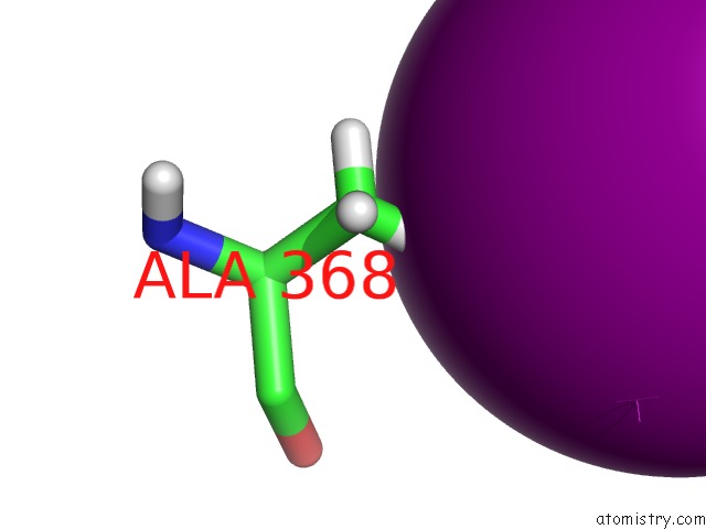
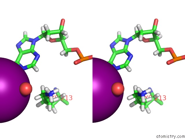
Iodine binding site 1 out of 9 in 5td5
Go back to
Iodine binding site 1 out
of 9 in the Crystal Structure of Human APOBEC3B Variant Complexed with Ssdna
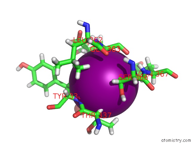
Mono view

Stereo pair view
Mono view
Stereo pair view
A full contact list of Iodine with other atoms in the I binding
site number 1 of Crystal Structure of Human APOBEC3B Variant Complexed with Ssdna within 5.0Å range:
|
Iodine binding site 2 out of 9 in 5td5
Go back to
Iodine binding site 2 out
of 9 in the Crystal Structure of Human APOBEC3B Variant Complexed with Ssdna

Mono view

Stereo pair view
Mono view
Stereo pair view
A full contact list of Iodine with other atoms in the I binding
site number 2 of Crystal Structure of Human APOBEC3B Variant Complexed with Ssdna within 5.0Å range:
|
Iodine binding site 3 out of 9 in 5td5
Go back to
Iodine binding site 3 out
of 9 in the Crystal Structure of Human APOBEC3B Variant Complexed with Ssdna

Mono view

Stereo pair view
Mono view
Stereo pair view
A full contact list of Iodine with other atoms in the I binding
site number 3 of Crystal Structure of Human APOBEC3B Variant Complexed with Ssdna within 5.0Å range:
|
Iodine binding site 4 out of 9 in 5td5
Go back to
Iodine binding site 4 out
of 9 in the Crystal Structure of Human APOBEC3B Variant Complexed with Ssdna

Mono view
Stereo pair view
Mono view
Stereo pair view
A full contact list of Iodine with other atoms in the I binding
site number 4 of Crystal Structure of Human APOBEC3B Variant Complexed with Ssdna within 5.0Å range:
|
Iodine binding site 5 out of 9 in 5td5
Go back to
Iodine binding site 5 out
of 9 in the Crystal Structure of Human APOBEC3B Variant Complexed with Ssdna

Mono view

Stereo pair view
Mono view
Stereo pair view
A full contact list of Iodine with other atoms in the I binding
site number 5 of Crystal Structure of Human APOBEC3B Variant Complexed with Ssdna within 5.0Å range:
|
Iodine binding site 6 out of 9 in 5td5
Go back to
Iodine binding site 6 out
of 9 in the Crystal Structure of Human APOBEC3B Variant Complexed with Ssdna
Mono view
Stereo pair view
Mono view
Stereo pair view
A full contact list of Iodine with other atoms in the I binding
site number 6 of Crystal Structure of Human APOBEC3B Variant Complexed with Ssdna within 5.0Å range:
|
Iodine binding site 7 out of 9 in 5td5
Go back to
Iodine binding site 7 out
of 9 in the Crystal Structure of Human APOBEC3B Variant Complexed with Ssdna
Mono view

Stereo pair view
Mono view
Stereo pair view
A full contact list of Iodine with other atoms in the I binding
site number 7 of Crystal Structure of Human APOBEC3B Variant Complexed with Ssdna within 5.0Å range:
|
Iodine binding site 8 out of 9 in 5td5
Go back to
Iodine binding site 8 out
of 9 in the Crystal Structure of Human APOBEC3B Variant Complexed with Ssdna

Mono view

Stereo pair view
Mono view
Stereo pair view
A full contact list of Iodine with other atoms in the I binding
site number 8 of Crystal Structure of Human APOBEC3B Variant Complexed with Ssdna within 5.0Å range:
|
Iodine binding site 9 out of 9 in 5td5
Go back to
Iodine binding site 9 out
of 9 in the Crystal Structure of Human APOBEC3B Variant Complexed with Ssdna

Mono view
Stereo pair view
Mono view
Stereo pair view
A full contact list of Iodine with other atoms in the I binding
site number 9 of Crystal Structure of Human APOBEC3B Variant Complexed with Ssdna within 5.0Å range:
|
Reference:
K.Shi,
M.A.Carpenter,
S.Banerjee,
N.M.Shaban,
K.Kurahashi,
D.J.Salamango,
J.L.Mccann,
G.J.Starrett,
J.V.Duffy,
O.Demir,
R.E.Amaro,
D.A.Harki,
R.S.Harris,
H.Aihara.
Structural Basis For Targeted Dna Cytosine Deamination and Mutagenesis By APOBEC3A and APOBEC3B. Nat. Struct. Mol. Biol. V. 24 131 2017.
ISSN: ESSN 1545-9985
PubMed: 27991903
DOI: 10.1038/NSMB.3344
Page generated: Sun Aug 11 21:52:41 2024
ISSN: ESSN 1545-9985
PubMed: 27991903
DOI: 10.1038/NSMB.3344
Last articles
Fe in 9IUYFe in 9IY1
Fe in 9J1I
Fe in 9J0J
Fe in 9ISU
Fe in 9IT8
Fe in 9ISS
Fe in 9IST
Fe in 9IS4
Fe in 9IOX