Iodine »
PDB 5w4i-6axx »
5zh9 »
Iodine in PDB 5zh9: Crystal Structures of Mutant Endo-Beta-1,4-Xylanase II (Y88F)
Enzymatic activity of Crystal Structures of Mutant Endo-Beta-1,4-Xylanase II (Y88F)
All present enzymatic activity of Crystal Structures of Mutant Endo-Beta-1,4-Xylanase II (Y88F):
3.2.1.8;
3.2.1.8;
Protein crystallography data
The structure of Crystal Structures of Mutant Endo-Beta-1,4-Xylanase II (Y88F), PDB code: 5zh9
was solved by
X.Zhang,
Q.Wan,
with X-Ray Crystallography technique. A brief refinement statistics is given in the table below:
| Resolution Low / High (Å) | 39.78 / 1.15 |
| Space group | P 21 21 21 |
| Cell size a, b, c (Å), α, β, γ (°) | 48.331, 59.327, 70.056, 90.00, 90.00, 90.00 |
| R / Rfree (%) | 13.7 / 14.8 |
Iodine Binding Sites:
The binding sites of Iodine atom in the Crystal Structures of Mutant Endo-Beta-1,4-Xylanase II (Y88F)
(pdb code 5zh9). This binding sites where shown within
5.0 Angstroms radius around Iodine atom.
In total 3 binding sites of Iodine where determined in the Crystal Structures of Mutant Endo-Beta-1,4-Xylanase II (Y88F), PDB code: 5zh9:
Jump to Iodine binding site number: 1; 2; 3;
In total 3 binding sites of Iodine where determined in the Crystal Structures of Mutant Endo-Beta-1,4-Xylanase II (Y88F), PDB code: 5zh9:
Jump to Iodine binding site number: 1; 2; 3;
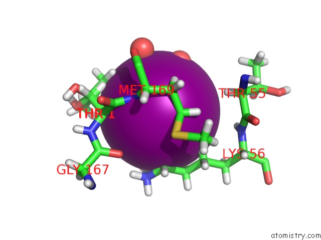
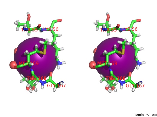
Iodine binding site 1 out of 3 in 5zh9
Go back to
Iodine binding site 1 out
of 3 in the Crystal Structures of Mutant Endo-Beta-1,4-Xylanase II (Y88F)
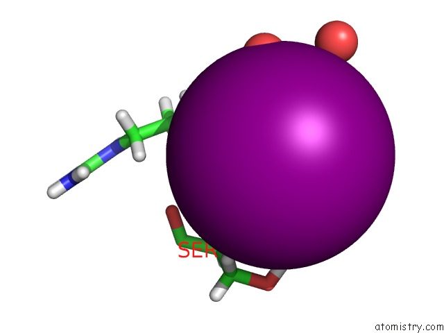
Mono view
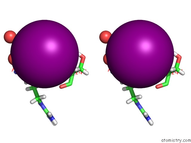
Stereo pair view
Mono view
Stereo pair view
A full contact list of Iodine with other atoms in the I binding
site number 1 of Crystal Structures of Mutant Endo-Beta-1,4-Xylanase II (Y88F) within 5.0Å range:
|
Iodine binding site 2 out of 3 in 5zh9
Go back to
Iodine binding site 2 out
of 3 in the Crystal Structures of Mutant Endo-Beta-1,4-Xylanase II (Y88F)
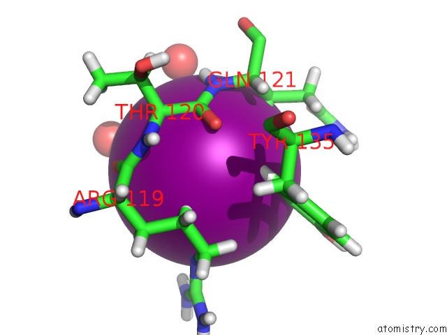
Mono view
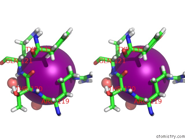
Stereo pair view
Mono view
Stereo pair view
A full contact list of Iodine with other atoms in the I binding
site number 2 of Crystal Structures of Mutant Endo-Beta-1,4-Xylanase II (Y88F) within 5.0Å range:
|
Iodine binding site 3 out of 3 in 5zh9
Go back to
Iodine binding site 3 out
of 3 in the Crystal Structures of Mutant Endo-Beta-1,4-Xylanase II (Y88F)
Mono view
Stereo pair view
Mono view
Stereo pair view
A full contact list of Iodine with other atoms in the I binding
site number 3 of Crystal Structures of Mutant Endo-Beta-1,4-Xylanase II (Y88F) within 5.0Å range:
|
Reference:
X.Zhang,
Q.Wan.
Crystal Structures of Mutant Endo-Beta-1,4-Xylanase II To Be Published.
Page generated: Sun Aug 11 22:25:23 2024
Last articles
I in 5ENMI in 5ENK
I in 5EEK
I in 5E9O
I in 5EMS
I in 5EIJ
I in 5EI1
I in 5DLV
I in 5EHB
I in 5E5Y