Atomistry » Iodine » PDB 6k9o-6nfk » 6kwg
Atomistry »
  Iodine »
    PDB 6k9o-6nfk »
      6kwg »

Iodine in PDB 6kwg: Crystal Structure Analysis of Endo-Beta-1,4-Xylanase II Complexed with Xylotriose

Enzymatic activity of Crystal Structure Analysis of Endo-Beta-1,4-Xylanase II Complexed with Xylotriose

All present enzymatic activity of Crystal Structure Analysis of Endo-Beta-1,4-Xylanase II Complexed with Xylotriose:
3.2.1.8;

Protein crystallography data

The structure of Crystal Structure Analysis of Endo-Beta-1,4-Xylanase II Complexed with Xylotriose, PDB code: 6kwg was solved by C.Li, Q.Wan, with X-Ray Crystallography technique. A brief refinement statistics is given in the table below:

Resolution Low / High (Å) 19.50 / 1.69
Space group P 21 21 21
Cell size a, b, c (Å), α, β, γ (°) 46.915, 58.533, 69.138, 90, 90, 90
R / Rfree (%) 20.9 / 24.4

Iodine Binding Sites:

The binding sites of Iodine atom in the Crystal Structure Analysis of Endo-Beta-1,4-Xylanase II Complexed with Xylotriose (pdb code 6kwg). This binding sites where shown within 5.0 Angstroms radius around Iodine atom.
In total 7 binding sites of Iodine where determined in the Crystal Structure Analysis of Endo-Beta-1,4-Xylanase II Complexed with Xylotriose, PDB code: 6kwg:
Jump to Iodine binding site number: 1; 2; 3; 4; 5; 6; 7;

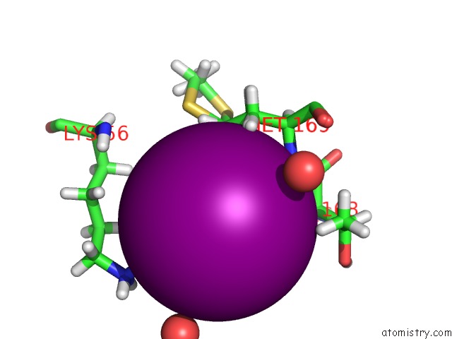
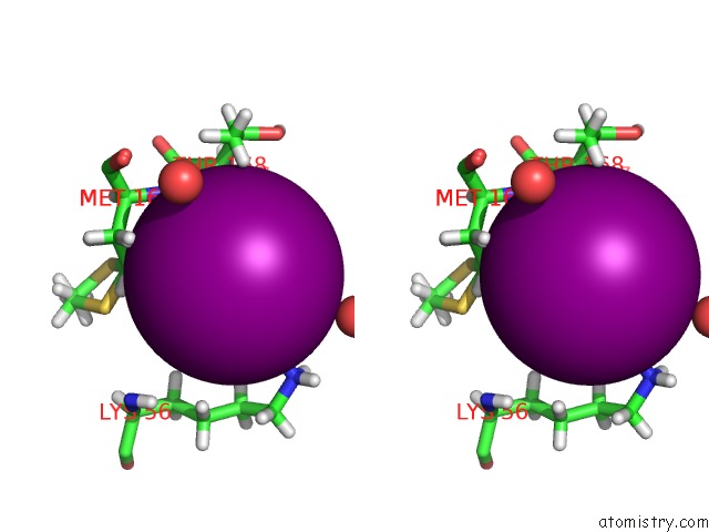
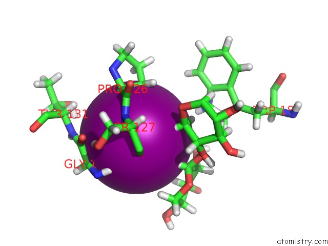
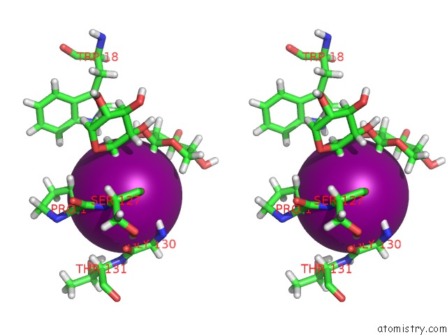
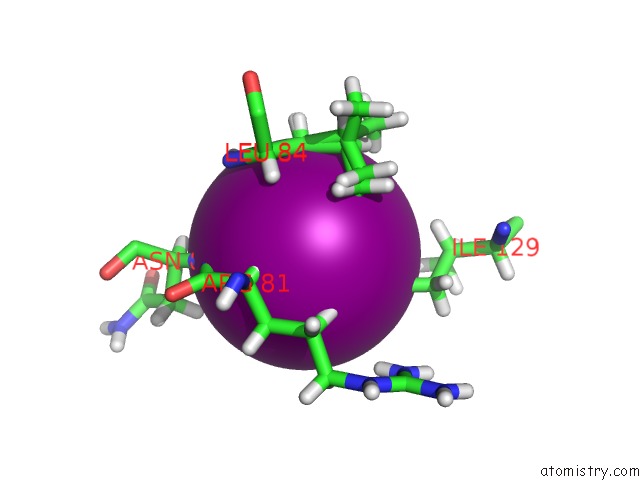
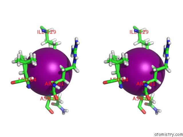
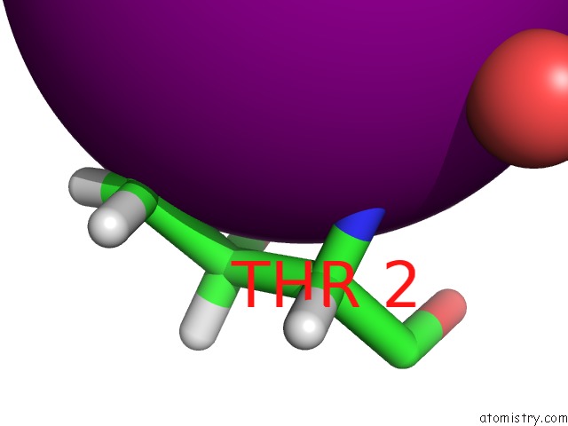
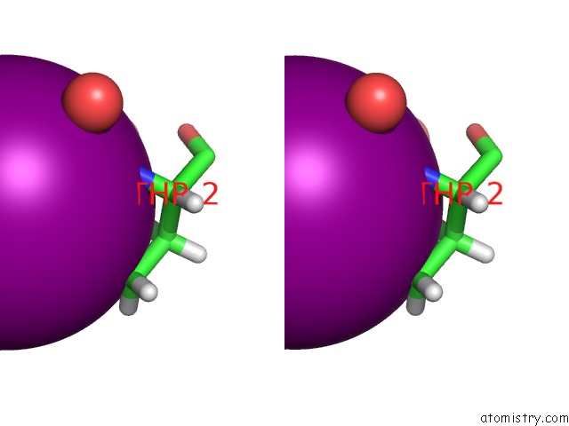
Iodine binding site 1 out of 7 in 6kwg

Go back to Iodine Binding Sites List in 6kwg
Iodine binding site 1 out of 7 in the Crystal Structure Analysis of Endo-Beta-1,4-Xylanase II Complexed with Xylotriose


Mono view


Stereo pair view

A full contact list of Iodine with other atoms in the I binding site number 1 of Crystal Structure Analysis of Endo-Beta-1,4-Xylanase II Complexed with Xylotriose within 5.0Å range:
probe atom residue distance (Å) B Occ
A:I204

b:166.2
occ:1.00
HG3 A:MET169 2.9 39.7 0.3
HG2 A:MET169 2.9 40.0 0.7
H A:MET169 3.1 37.0 0.7
H A:MET169 3.1 37.1 0.3
HA A:THR168 3.4 36.3 1.0
HZ3 A:LYS56 3.5 68.1 1.0
CG A:MET169 3.8 33.3 0.7
CG A:MET169 3.8 33.0 0.3
N A:MET169 3.9 30.8 0.7
N A:MET169 3.9 30.9 0.3
HB2 A:MET169 4.1 36.1 0.3
HG3 A:MET169 4.1 40.0 0.7
HB2 A:MET169 4.1 36.0 0.7
O A:GLY167 4.2 32.1 1.0
HG2 A:MET169 4.2 39.7 0.3
HG23 A:THR168 4.2 46.0 1.0
CA A:THR168 4.3 30.2 1.0
NZ A:LYS56 4.3 56.7 1.0
HD3 A:LYS56 4.4 58.3 1.0
CB A:MET169 4.4 30.1 0.3
CB A:MET169 4.4 30.0 0.7
HZ2 A:LYS56 4.5 68.1 1.0
C A:THR168 4.6 28.6 1.0
O A:HOH458 4.6 47.3 1.0
HZ1 A:LYS56 4.7 68.1 1.0
HB3 A:LYS56 4.7 42.9 1.0
CA A:MET169 4.8 29.0 0.7
CA A:MET169 4.8 29.4 0.3
SD A:MET169 4.9 36.1 0.3
HG2 A:LYS56 4.9 54.5 1.0
O A:HOH344 4.9 39.2 1.0
CG2 A:THR168 5.0 38.3 1.0

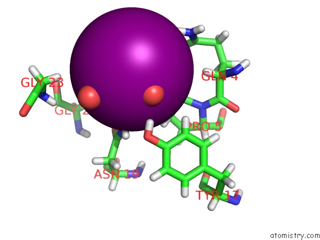
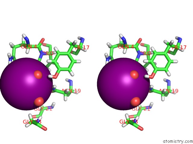
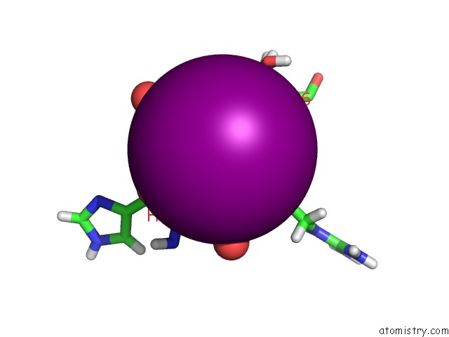
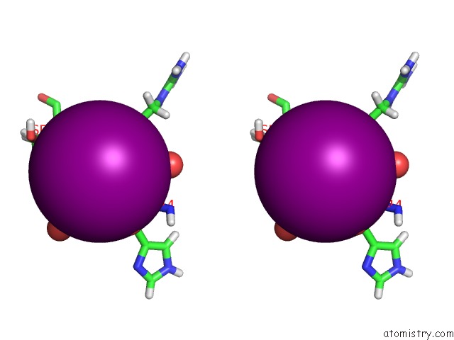
Iodine binding site 2 out of 7 in 6kwg

Go back to Iodine Binding Sites List in 6kwg
Iodine binding site 2 out of 7 in the Crystal Structure Analysis of Endo-Beta-1,4-Xylanase II Complexed with Xylotriose


Mono view


Stereo pair view

A full contact list of Iodine with other atoms in the I binding site number 2 of Crystal Structure Analysis of Endo-Beta-1,4-Xylanase II Complexed with Xylotriose within 5.0Å range:
probe atom residue distance (Å) B Occ
A:I205

b:64.6
occ:1.00
HE A:ARG119 3.2 50.1 1.0
HE1 A:TYR135 3.3 35.5 1.0
HA A:THR120 3.3 34.1 1.0
HG2 A:ARG119 3.5 34.0 1.0
HB2 A:GLN121 3.6 36.6 1.0
HD1 A:TYR135 3.7 34.5 1.0
O A:ARG119 3.9 27.0 1.0
C A:THR120 4.0 28.1 1.0
CA A:THR120 4.0 28.4 1.0
CE1 A:TYR135 4.0 29.6 1.0
NE A:ARG119 4.0 41.7 1.0
O A:HOH331 4.0 50.2 1.0
C A:ARG119 4.2 23.3 1.0
O A:HOH383 4.2 36.9 1.0
N A:GLN121 4.2 31.2 1.0
CD1 A:TYR135 4.2 28.7 1.0
CG A:ARG119 4.3 28.3 1.0
HB3 A:ARG119 4.3 35.3 1.0
O A:THR120 4.3 26.6 1.0
N A:THR120 4.3 24.4 1.0
CB A:GLN121 4.4 30.5 1.0
H A:GLN121 4.4 37.4 1.0
HB3 A:GLN121 4.4 36.6 1.0
O A:HOH480 4.4 46.8 1.0
HD2 A:ARG119 4.5 39.5 1.0
HH21 A:ARG119 4.5 51.9 1.0
CD A:ARG119 4.5 32.9 1.0
O A:HOH450 4.7 36.9 1.0
CB A:ARG119 4.8 29.4 1.0
O A:HOH301 4.9 42.4 1.0
CA A:GLN121 4.9 27.6 1.0
H A:THR120 5.0 29.3 1.0

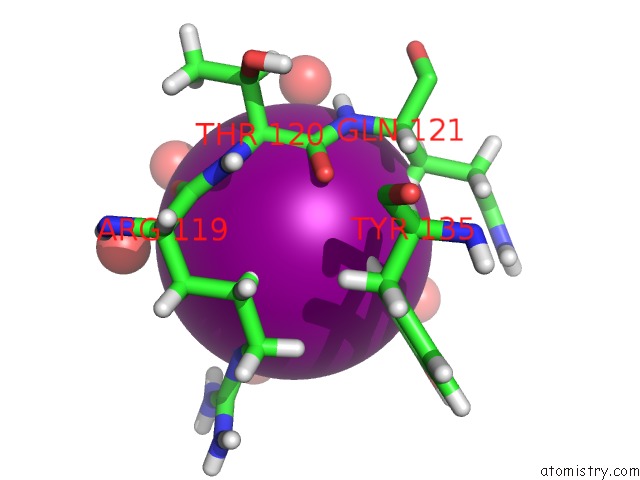
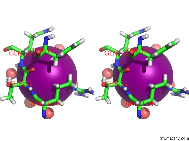
Iodine binding site 3 out of 7 in 6kwg

Go back to Iodine Binding Sites List in 6kwg
Iodine binding site 3 out of 7 in the Crystal Structure Analysis of Endo-Beta-1,4-Xylanase II Complexed with Xylotriose


Mono view


Stereo pair view

A full contact list of Iodine with other atoms in the I binding site number 3 of Crystal Structure Analysis of Endo-Beta-1,4-Xylanase II Complexed with Xylotriose within 5.0Å range:
probe atom residue distance (Å) B Occ
A:I206

b:65.7
occ:1.00
HB3 A:PRO126 3.0 43.4 1.0
O A:HOH474 3.1 37.5 1.0
HA2 A:GLY130 3.2 30.5 1.0
H A:SER127 3.3 35.2 1.0
HA A:PRO126 3.4 39.1 1.0
O A:SER127 3.5 27.9 1.0
H1 B:XYP3 3.5 41.9 1.0
H51 B:XYP2 3.6 36.7 1.0
CB A:PRO126 3.9 36.1 1.0
H A:GLY130 3.9 34.8 1.0
N A:SER127 3.9 29.3 1.0
CA A:PRO126 4.0 32.6 1.0
CA A:GLY130 4.0 25.4 1.0
H3 B:XYP3 4.2 59.3 1.0
C A:GLY130 4.2 28.2 1.0
O2 B:XYP3 4.3 41.4 1.0
HB2 A:PRO126 4.4 43.4 1.0
N A:GLY130 4.4 29.0 1.0
C1 B:XYP3 4.4 34.9 1.0
C5 B:XYP2 4.4 30.6 1.0
H52 B:XYP2 4.5 36.7 1.0
C A:PRO126 4.5 31.0 1.0
N A:THR131 4.5 27.1 1.0
H A:THR131 4.5 32.6 1.0
C A:SER127 4.6 27.6 1.0
O A:GLY130 4.7 27.4 1.0
HG3 A:PRO126 4.7 40.6 1.0
C2 B:XYP3 4.7 34.5 1.0
HE1 A:TRP18 4.7 40.1 1.0
H51 B:XYP3 4.8 42.4 1.0
HA3 A:GLY130 4.8 30.5 1.0
HG1 A:THR131 4.8 38.3 1.0
CA A:SER127 4.9 28.5 1.0
C3 B:XYP3 4.9 49.4 1.0
CG A:PRO126 4.9 33.8 1.0

Iodine binding site 4 out of 7 in 6kwg

Go back to Iodine Binding Sites List in 6kwg
Iodine binding site 4 out of 7 in the Crystal Structure Analysis of Endo-Beta-1,4-Xylanase II Complexed with Xylotriose


Mono view


Stereo pair view

A full contact list of Iodine with other atoms in the I binding site number 4 of Crystal Structure Analysis of Endo-Beta-1,4-Xylanase II Complexed with Xylotriose within 5.0Å range:
probe atom residue distance (Å) B Occ
A:I207

b:65.4
occ:1.00
HD21 A:ASN19 3.0 45.0 1.0
HD3 A:PRO5 3.3 46.3 1.0
O A:HOH342 3.6 36.1 1.0
HD2 A:PRO5 3.8 46.3 1.0
ND2 A:ASN19 3.8 37.5 1.0
HE22 A:GLN4 3.8 38.0 1.0
CD A:PRO5 3.9 38.6 1.0
HG2 A:PRO5 3.9 44.5 1.0
O A:HOH464 4.0 35.1 1.0
HG3 A:PRO5 4.2 44.5 1.0
OE1 A:GLN4 4.2 36.3 1.0
CG A:PRO5 4.2 37.0 1.0
HD22 A:ASN19 4.3 45.0 1.0
NE2 A:GLN4 4.5 31.7 1.0
HH A:TYR17 4.5 42.0 1.0
OD1 A:ASN19 4.6 37.8 1.0
CG A:ASN19 4.7 37.5 1.0
CD A:GLN4 4.7 32.9 1.0
HA A:GLN4 4.8 39.4 1.0
HA2 A:GLY23 5.0 51.7 1.0
HA2 A:GLY21 5.0 54.6 1.0

Iodine binding site 5 out of 7 in 6kwg

Go back to Iodine Binding Sites List in 6kwg
Iodine binding site 5 out of 7 in the Crystal Structure Analysis of Endo-Beta-1,4-Xylanase II Complexed with Xylotriose


Mono view


Stereo pair view

A full contact list of Iodine with other atoms in the I binding site number 5 of Crystal Structure Analysis of Endo-Beta-1,4-Xylanase II Complexed with Xylotriose within 5.0Å range:
probe atom residue distance (Å) B Occ
A:I208

b:26.0
occ:1.00
H A:ASN82 2.8 26.4 1.0
HD13 A:ILE129 2.8 49.8 1.0
HD12 A:ILE129 2.9 49.8 1.0
HB2 A:LEU84 3.1 29.2 0.0
HB2 A:LEU84 3.1 30.1 1.0
HD23 A:LEU84 3.2 29.6 0.0
CD1 A:ILE129 3.2 41.5 1.0
HB3 A:ARG81 3.5 28.1 1.0
HD11 A:ILE129 3.5 49.8 1.0
HA A:ARG81 3.5 30.2 1.0
N A:ASN82 3.5 22.0 1.0
HD13 A:LEU84 3.5 29.8 1.0
HG2 A:ARG81 3.8 31.9 1.0
CB A:LEU84 4.0 24.3 0.0
CB A:LEU84 4.0 25.1 1.0
CD2 A:LEU84 4.1 24.6 0.0
CB A:ARG81 4.1 23.4 1.0
CA A:ARG81 4.1 25.2 1.0
H A:LEU84 4.2 31.4 1.0
HA A:ASN82 4.2 33.5 1.0
H A:LEU84 4.2 30.5 0.0
HB2 A:ASN82 4.2 28.9 1.0
HG21 A:ILE129 4.2 47.4 1.0
HA A:LEU84 4.2 30.3 1.0
HA A:LEU84 4.2 29.6 0.0
C A:ARG81 4.3 27.1 1.0
HD22 A:LEU84 4.4 31.5 1.0
CD1 A:LEU84 4.4 24.8 1.0
N A:LEU84 4.4 26.2 1.0
N A:LEU84 4.4 25.4 0.0
CA A:ASN82 4.4 27.9 1.0
HD22 A:LEU84 4.4 29.6 0.0
HG A:LEU84 4.4 31.3 0.0
CG A:LEU84 4.4 26.0 0.0
CA A:LEU84 4.5 24.6 0.0
CG A:ARG81 4.5 26.6 1.0
CA A:LEU84 4.5 25.2 1.0
HD12 A:LEU84 4.6 29.8 1.0
CG1 A:ILE129 4.6 31.0 1.0
HB A:ILE129 4.6 37.7 1.0
HB3 A:LEU84 4.6 30.1 1.0
HB3 A:LEU84 4.7 29.2 0.0
CG A:LEU84 4.7 25.5 1.0
HD21 A:LEU84 4.8 29.6 0.0
CB A:ASN82 4.8 24.1 1.0
HH11 A:ARG81 4.9 44.2 1.0
HD3 A:ARG81 5.0 36.6 1.0
HB2 A:ARG81 5.0 28.1 1.0
CB A:ILE129 5.0 31.4 1.0

Iodine binding site 6 out of 7 in 6kwg

Go back to Iodine Binding Sites List in 6kwg
Iodine binding site 6 out of 7 in the Crystal Structure Analysis of Endo-Beta-1,4-Xylanase II Complexed with Xylotriose


Mono view


Stereo pair view

A full contact list of Iodine with other atoms in the I binding site number 6 of Crystal Structure Analysis of Endo-Beta-1,4-Xylanase II Complexed with Xylotriose within 5.0Å range:
probe atom residue distance (Å) B Occ
A:I209

b:142.5
occ:1.00
O A:HOH502 2.7 36.3 1.0
HA A:ARG145 2.8 31.9 1.0
O A:HIS144 2.8 26.6 1.0
O A:HOH466 3.3 51.2 1.0
CA A:ARG145 3.7 26.6 1.0
O A:HOH315 3.7 31.3 1.0
C A:HIS144 3.7 27.4 1.0
O A:HOH349 3.9 35.6 1.0
HB3 A:ARG145 4.0 28.6 1.0
N A:ARG145 4.1 25.7 1.0
CB A:ARG145 4.2 23.8 1.0
HB2 A:ARG145 4.3 28.6 1.0
H A:SER146 4.3 29.4 1.0
O A:HOH354 4.6 37.4 1.0
HB2 A:HIS144 4.7 33.7 1.0
C A:ARG145 4.7 24.6 1.0
N A:SER146 4.8 24.5 1.0
HD21 A:ASN92 4.8 35.6 1.0
H A:ARG145 4.9 30.9 1.0
CA A:HIS144 5.0 26.9 1.0

Iodine binding site 7 out of 7 in 6kwg

Go back to Iodine Binding Sites List in 6kwg
Iodine binding site 7 out of 7 in the Crystal Structure Analysis of Endo-Beta-1,4-Xylanase II Complexed with Xylotriose


Mono view


Stereo pair view

A full contact list of Iodine with other atoms in the I binding site number 7 of Crystal Structure Analysis of Endo-Beta-1,4-Xylanase II Complexed with Xylotriose within 5.0Å range:
probe atom residue distance (Å) B Occ
A:I210

b:86.8
occ:1.00
HG23 A:THR2 3.1 54.2 1.0
N A:THR2 3.6 45.6 1.0
O A:HOH312 3.7 46.7 1.0
OG1 A:THR2 3.8 47.4 1.0
CG2 A:THR2 4.0 45.1 1.0
HG1 A:THR2 4.0 56.9 1.0
O A:HOH305 4.3 51.9 1.0
HG21 A:THR2 4.3 54.2 1.0
CB A:THR2 4.4 37.5 1.0
CA A:THR2 4.6 40.4 1.0
HG22 A:THR2 4.7 54.2 1.0

Reference:

Z.Li, X.Zhang, C.Li, A.Kovalevsky, Q.Wan. Studying the Role of A Single Mutation of A Family 11 Glycoside Hydrolase Using High-Resolution X-Ray Crystallography. Protein J. V. 39 671 2020.
ISSN: ISSN 1572-3887
PubMed: 33128114
DOI: 10.1007/S10930-020-09938-5
Page generated: Sun Aug 11 23:30:20 2024

Last articles

I in 5EUZ
I in 5ENM
I in 5ENK
I in 5EEK
I in 5E9O
I in 5EMS
I in 5EIJ
I in 5EI1
I in 5DLV
I in 5EHB
© Copyright 2008-2020 by atomistry.com
Home   |    Site Map   |    Copyright   |    Contact us   |    Privacy