Iodine »
PDB 7lxr-7px1 »
7pcp »
Iodine in PDB 7pcp: Crystal Structure of CD73 in Complex with 5-Iodouracil in the Open Form
Enzymatic activity of Crystal Structure of CD73 in Complex with 5-Iodouracil in the Open Form
All present enzymatic activity of Crystal Structure of CD73 in Complex with 5-Iodouracil in the Open Form:
3.1.3.5;
3.1.3.5;
Protein crystallography data
The structure of Crystal Structure of CD73 in Complex with 5-Iodouracil in the Open Form, PDB code: 7pcp
was solved by
E.R.Scaletti,
N.Strater,
with X-Ray Crystallography technique. A brief refinement statistics is given in the table below:
| Resolution Low / High (Å) | 47.26 / 1.38 |
| Space group | P 21 21 2 |
| Cell size a, b, c (Å), α, β, γ (°) | 67.22, 131.82, 66.324, 90, 90, 90 |
| R / Rfree (%) | 12.5 / 16.6 |
Other elements in 7pcp:
The structure of Crystal Structure of CD73 in Complex with 5-Iodouracil in the Open Form also contains other interesting chemical elements:
| Calcium | (Ca) | 1 atom |
| Zinc | (Zn) | 2 atoms |
Iodine Binding Sites:
The binding sites of Iodine atom in the Crystal Structure of CD73 in Complex with 5-Iodouracil in the Open Form
(pdb code 7pcp). This binding sites where shown within
5.0 Angstroms radius around Iodine atom.
In total only one binding site of Iodine was determined in the Crystal Structure of CD73 in Complex with 5-Iodouracil in the Open Form, PDB code: 7pcp:
In total only one binding site of Iodine was determined in the Crystal Structure of CD73 in Complex with 5-Iodouracil in the Open Form, PDB code: 7pcp:
Iodine binding site 1 out of 1 in 7pcp
Go back to
Iodine binding site 1 out
of 1 in the Crystal Structure of CD73 in Complex with 5-Iodouracil in the Open Form
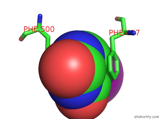
Mono view
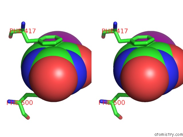
Stereo pair view
Mono view
Stereo pair view
A full contact list of Iodine with other atoms in the I binding
site number 1 of Crystal Structure of CD73 in Complex with 5-Iodouracil in the Open Form within 5.0Å range:
|
Reference:
E.Scaletti,
F.U.Huschmann,
U.Mueller,
M.S.Weiss,
N.Strater.
Substrate Binding Modes of Purine and Pyrimidine Nucleotides to Human Ecto-5'-Nucleotidase (CD73) and Inhibition By Their Bisphosphonic Acid Derivatives. Purinergic Signal 2021.
ISSN: ISSN 1573-9546
PubMed: 34403084
DOI: 10.1007/S11302-021-09802-W
Page generated: Fri Aug 8 23:32:49 2025
ISSN: ISSN 1573-9546
PubMed: 34403084
DOI: 10.1007/S11302-021-09802-W
Last articles
Mn in 4X4OMn in 4XJK
Mn in 4XIS
Mn in 4XBW
Mn in 4X9Q
Mn in 4WV8
Mn in 4X8D
Mn in 4WZQ
Mn in 4WZM
Mn in 4WYL