Iodine »
PDB 7sia-8a24 »
7up5 »
Iodine in PDB 7up5: Crystal Structure of C-Terminal Domain of MSK1 in Complex with Covalently Bound Pyrrolopyrimidine Compound 23 (Co-Crystal)
Enzymatic activity of Crystal Structure of C-Terminal Domain of MSK1 in Complex with Covalently Bound Pyrrolopyrimidine Compound 23 (Co-Crystal)
All present enzymatic activity of Crystal Structure of C-Terminal Domain of MSK1 in Complex with Covalently Bound Pyrrolopyrimidine Compound 23 (Co-Crystal):
2.7.11.1;
2.7.11.1;
Protein crystallography data
The structure of Crystal Structure of C-Terminal Domain of MSK1 in Complex with Covalently Bound Pyrrolopyrimidine Compound 23 (Co-Crystal), PDB code: 7up5
was solved by
J.K.Yano,
T.E.Edwards,
A.Hall,
with X-Ray Crystallography technique. A brief refinement statistics is given in the table below:
| Resolution Low / High (Å) | 48.15 / 2.80 |
| Space group | P 21 21 21 |
| Cell size a, b, c (Å), α, β, γ (°) | 51.48, 90.78, 135.97, 90, 90, 90 |
| R / Rfree (%) | 20.1 / 26.8 |
Iodine Binding Sites:
The binding sites of Iodine atom in the Crystal Structure of C-Terminal Domain of MSK1 in Complex with Covalently Bound Pyrrolopyrimidine Compound 23 (Co-Crystal)
(pdb code 7up5). This binding sites where shown within
5.0 Angstroms radius around Iodine atom.
In total 3 binding sites of Iodine where determined in the Crystal Structure of C-Terminal Domain of MSK1 in Complex with Covalently Bound Pyrrolopyrimidine Compound 23 (Co-Crystal), PDB code: 7up5:
Jump to Iodine binding site number: 1; 2; 3;
In total 3 binding sites of Iodine where determined in the Crystal Structure of C-Terminal Domain of MSK1 in Complex with Covalently Bound Pyrrolopyrimidine Compound 23 (Co-Crystal), PDB code: 7up5:
Jump to Iodine binding site number: 1; 2; 3;
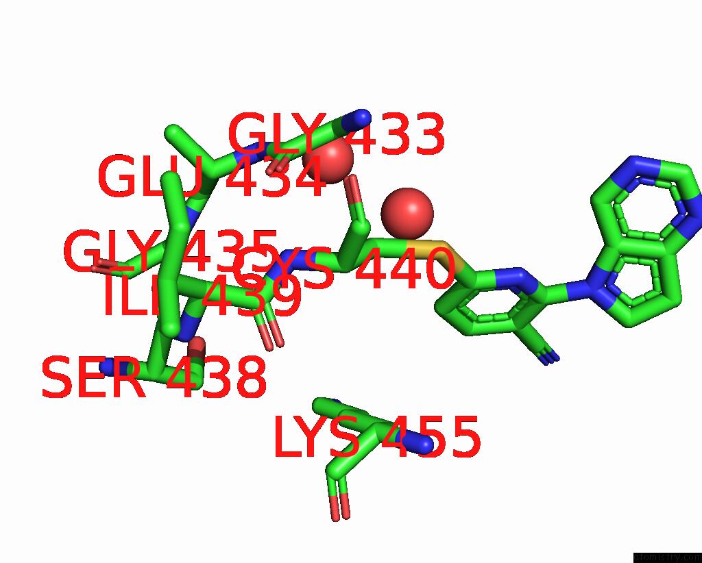
Iodine binding site 1 out of 3 in 7up5
Go back to
Iodine binding site 1 out
of 3 in the Crystal Structure of C-Terminal Domain of MSK1 in Complex with Covalently Bound Pyrrolopyrimidine Compound 23 (Co-Crystal)
Mono view
Stereo pair view
Mono view
Stereo pair view
A full contact list of Iodine with other atoms in the I binding
site number 1 of Crystal Structure of C-Terminal Domain of MSK1 in Complex with Covalently Bound Pyrrolopyrimidine Compound 23 (Co-Crystal) within 5.0Å range:
|
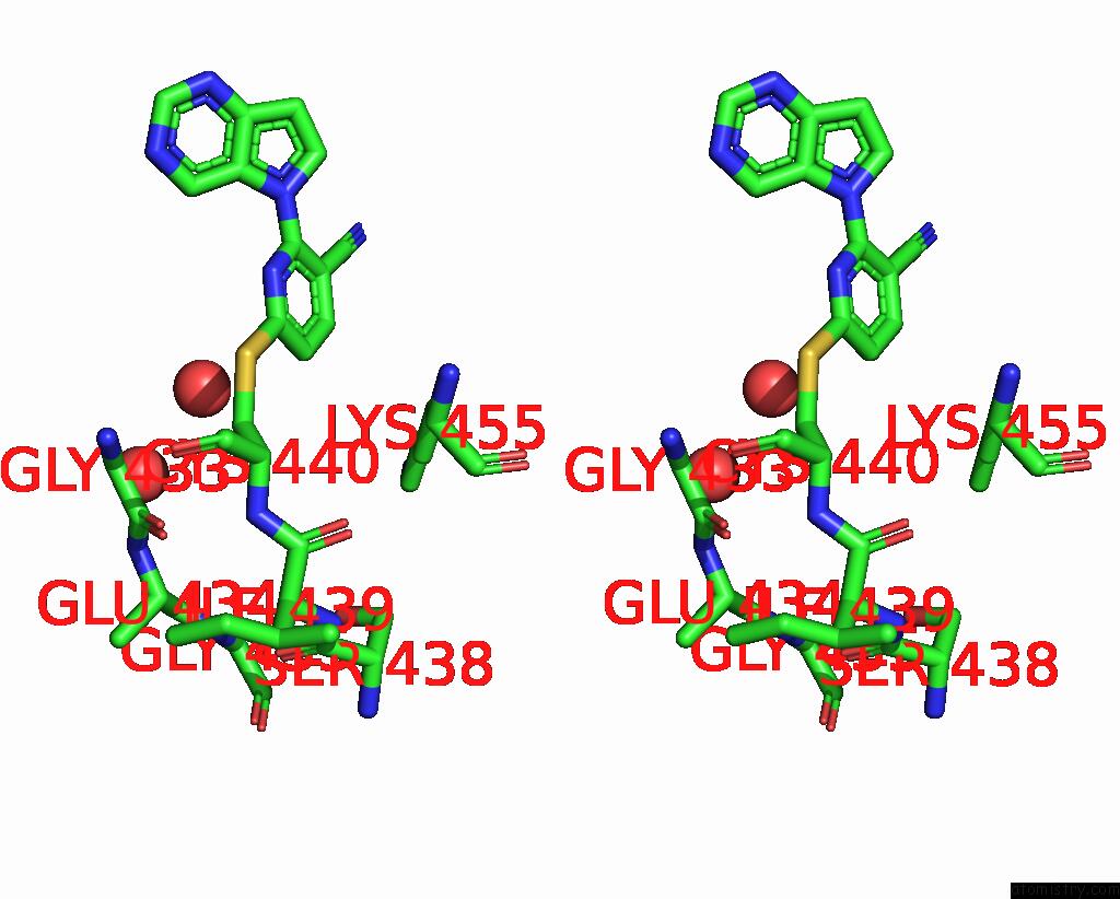
Iodine binding site 2 out of 3 in 7up5
Go back to
Iodine binding site 2 out
of 3 in the Crystal Structure of C-Terminal Domain of MSK1 in Complex with Covalently Bound Pyrrolopyrimidine Compound 23 (Co-Crystal)

Mono view

Stereo pair view
Mono view
Stereo pair view
A full contact list of Iodine with other atoms in the I binding
site number 2 of Crystal Structure of C-Terminal Domain of MSK1 in Complex with Covalently Bound Pyrrolopyrimidine Compound 23 (Co-Crystal) within 5.0Å range:
|
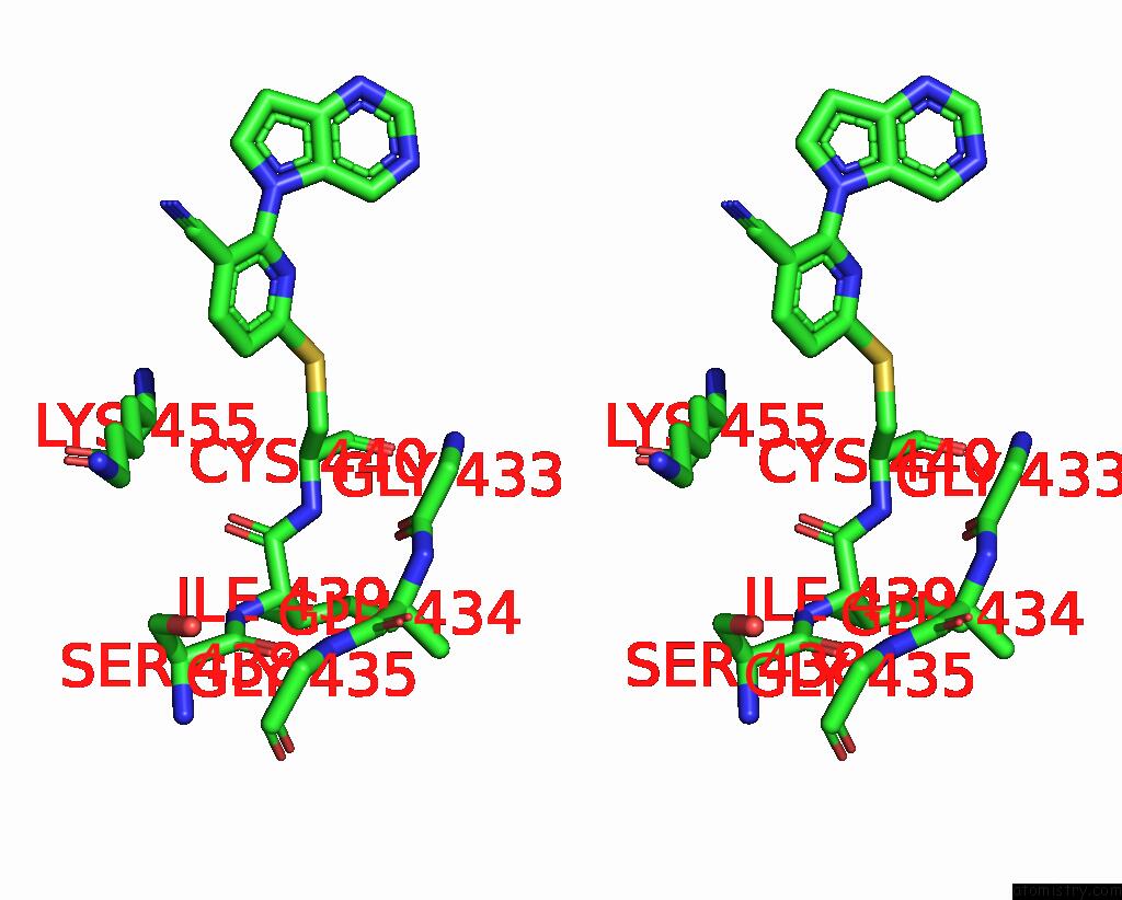
Iodine binding site 3 out of 3 in 7up5
Go back to
Iodine binding site 3 out
of 3 in the Crystal Structure of C-Terminal Domain of MSK1 in Complex with Covalently Bound Pyrrolopyrimidine Compound 23 (Co-Crystal)

Mono view
Stereo pair view
Mono view
Stereo pair view
A full contact list of Iodine with other atoms in the I binding
site number 3 of Crystal Structure of C-Terminal Domain of MSK1 in Complex with Covalently Bound Pyrrolopyrimidine Compound 23 (Co-Crystal) within 5.0Å range:
|
Reference:
A.Hall,
J.Abendroth,
M.J.Bolejack,
T.Ceska,
S.Dell'aiera,
V.Ellis,
D.Fox 3Rd,
C.Francois,
M.M.Muruthi,
C.Prevel,
K.Poullennec,
S.Romanov,
A.Valade,
A.Vanbellinghen,
J.Yano,
M.Geraerts.
Discovery and Characterization of A Novel Series of Chloropyrimidines As Covalent Inhibitors of the Kinase MSK1. Acs Med.Chem.Lett. V. 13 1099 2022.
ISSN: ISSN 1948-5875
PubMed: 35859861
DOI: 10.1021/ACSMEDCHEMLETT.2C00134
Page generated: Fri Aug 8 23:48:06 2025
ISSN: ISSN 1948-5875
PubMed: 35859861
DOI: 10.1021/ACSMEDCHEMLETT.2C00134
Last articles
Na in 6ZXZNa in 6ZZW
Na in 6ZYR
Na in 6ZS1
Na in 6ZXX
Na in 6ZWK
Na in 6ZXU
Na in 6ZUL
Na in 6ZXT
Na in 6ZUM