Iodine »
PDB 7sia-8a24 »
7xc1 »
Iodine in PDB 7xc1: Crystal Structure of ERK2 with An Allosteric Inhibitor 3
Enzymatic activity of Crystal Structure of ERK2 with An Allosteric Inhibitor 3
All present enzymatic activity of Crystal Structure of ERK2 with An Allosteric Inhibitor 3:
2.7.11.24;
2.7.11.24;
Protein crystallography data
The structure of Crystal Structure of ERK2 with An Allosteric Inhibitor 3, PDB code: 7xc1
was solved by
M.Yoshida,
T.Kinoshita,
with X-Ray Crystallography technique. A brief refinement statistics is given in the table below:
| Resolution Low / High (Å) | 41.33 / 2.09 |
| Space group | P 41 21 2 |
| Cell size a, b, c (Å), α, β, γ (°) | 82.653, 82.653, 275.074, 90, 90, 90 |
| R / Rfree (%) | 23 / 27.1 |
Other elements in 7xc1:
The structure of Crystal Structure of ERK2 with An Allosteric Inhibitor 3 also contains other interesting chemical elements:
| Fluorine | (F) | 1 atom |
Iodine Binding Sites:
The binding sites of Iodine atom in the Crystal Structure of ERK2 with An Allosteric Inhibitor 3
(pdb code 7xc1). This binding sites where shown within
5.0 Angstroms radius around Iodine atom.
In total 2 binding sites of Iodine where determined in the Crystal Structure of ERK2 with An Allosteric Inhibitor 3, PDB code: 7xc1:
Jump to Iodine binding site number: 1; 2;
In total 2 binding sites of Iodine where determined in the Crystal Structure of ERK2 with An Allosteric Inhibitor 3, PDB code: 7xc1:
Jump to Iodine binding site number: 1; 2;
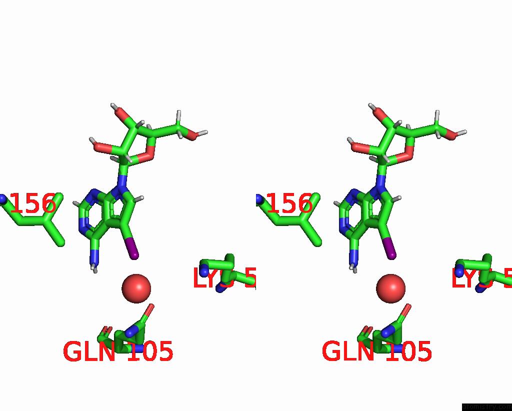
Iodine binding site 1 out of 2 in 7xc1
Go back to
Iodine binding site 1 out
of 2 in the Crystal Structure of ERK2 with An Allosteric Inhibitor 3
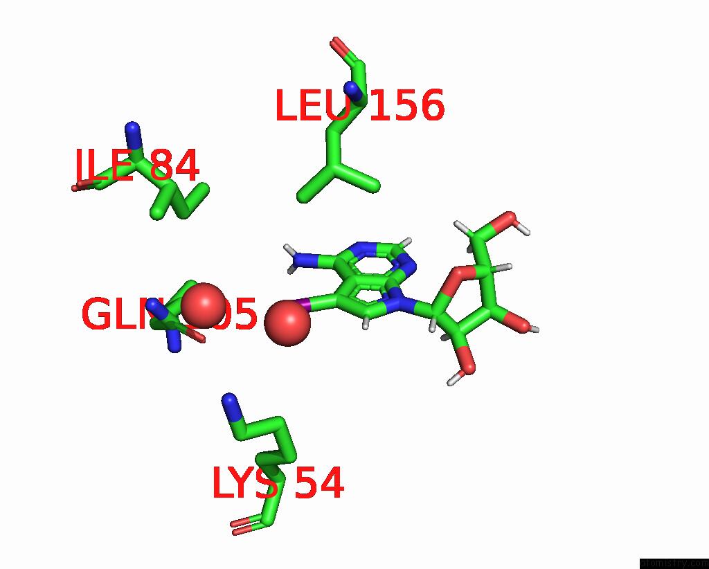
Mono view

Stereo pair view
Mono view
Stereo pair view
A full contact list of Iodine with other atoms in the I binding
site number 1 of Crystal Structure of ERK2 with An Allosteric Inhibitor 3 within 5.0Å range:
|
Iodine binding site 2 out of 2 in 7xc1
Go back to
Iodine binding site 2 out
of 2 in the Crystal Structure of ERK2 with An Allosteric Inhibitor 3

Mono view
Stereo pair view
Mono view
Stereo pair view
A full contact list of Iodine with other atoms in the I binding
site number 2 of Crystal Structure of ERK2 with An Allosteric Inhibitor 3 within 5.0Å range:
|
Reference:
M.Yoshida,
T.Kinoshita.
Structural Basis For ERK2 Allosteric Inhibitors. To Be Published.
Page generated: Fri Aug 8 23:53:24 2025
Last articles
Na in 5K8BNa in 5K8R
Na in 5K7O
Na in 5K4V
Na in 5K2D
Na in 5K2C
Na in 5K2B
Na in 5K0J
Na in 5K0G
Na in 5K2A