Iodine »
PDB 9axj-9rp9 »
9dq1 »
Iodine in PDB 9dq1: Crystal Structure of Hrmj From Streptomyces Sp. Cfmr 7 (Hrmj-Ssc) Complexed with Manganese (II), 2-Oxoglutarate and 6-Nitronorleucine
Protein crystallography data
The structure of Crystal Structure of Hrmj From Streptomyces Sp. Cfmr 7 (Hrmj-Ssc) Complexed with Manganese (II), 2-Oxoglutarate and 6-Nitronorleucine, PDB code: 9dq1
was solved by
Y.-C.Zheng,
P.Swartz,
W.-C.Chang,
with X-Ray Crystallography technique. A brief refinement statistics is given in the table below:
| Resolution Low / High (Å) | 17.49 / 1.90 |
| Space group | P 1 21 1 |
| Cell size a, b, c (Å), α, β, γ (°) | 44.342, 74.971, 87.295, 90, 90.03, 90 |
| R / Rfree (%) | 17.5 / 20.4 |
Other elements in 9dq1:
The structure of Crystal Structure of Hrmj From Streptomyces Sp. Cfmr 7 (Hrmj-Ssc) Complexed with Manganese (II), 2-Oxoglutarate and 6-Nitronorleucine also contains other interesting chemical elements:
| Manganese | (Mn) | 2 atoms |
Iodine Binding Sites:
The binding sites of Iodine atom in the Crystal Structure of Hrmj From Streptomyces Sp. Cfmr 7 (Hrmj-Ssc) Complexed with Manganese (II), 2-Oxoglutarate and 6-Nitronorleucine
(pdb code 9dq1). This binding sites where shown within
5.0 Angstroms radius around Iodine atom.
In total 7 binding sites of Iodine where determined in the Crystal Structure of Hrmj From Streptomyces Sp. Cfmr 7 (Hrmj-Ssc) Complexed with Manganese (II), 2-Oxoglutarate and 6-Nitronorleucine, PDB code: 9dq1:
Jump to Iodine binding site number: 1; 2; 3; 4; 5; 6; 7;
In total 7 binding sites of Iodine where determined in the Crystal Structure of Hrmj From Streptomyces Sp. Cfmr 7 (Hrmj-Ssc) Complexed with Manganese (II), 2-Oxoglutarate and 6-Nitronorleucine, PDB code: 9dq1:
Jump to Iodine binding site number: 1; 2; 3; 4; 5; 6; 7;
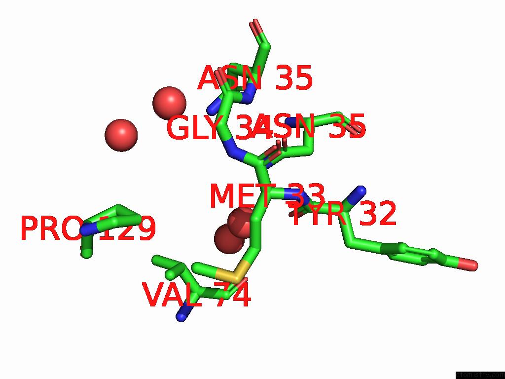
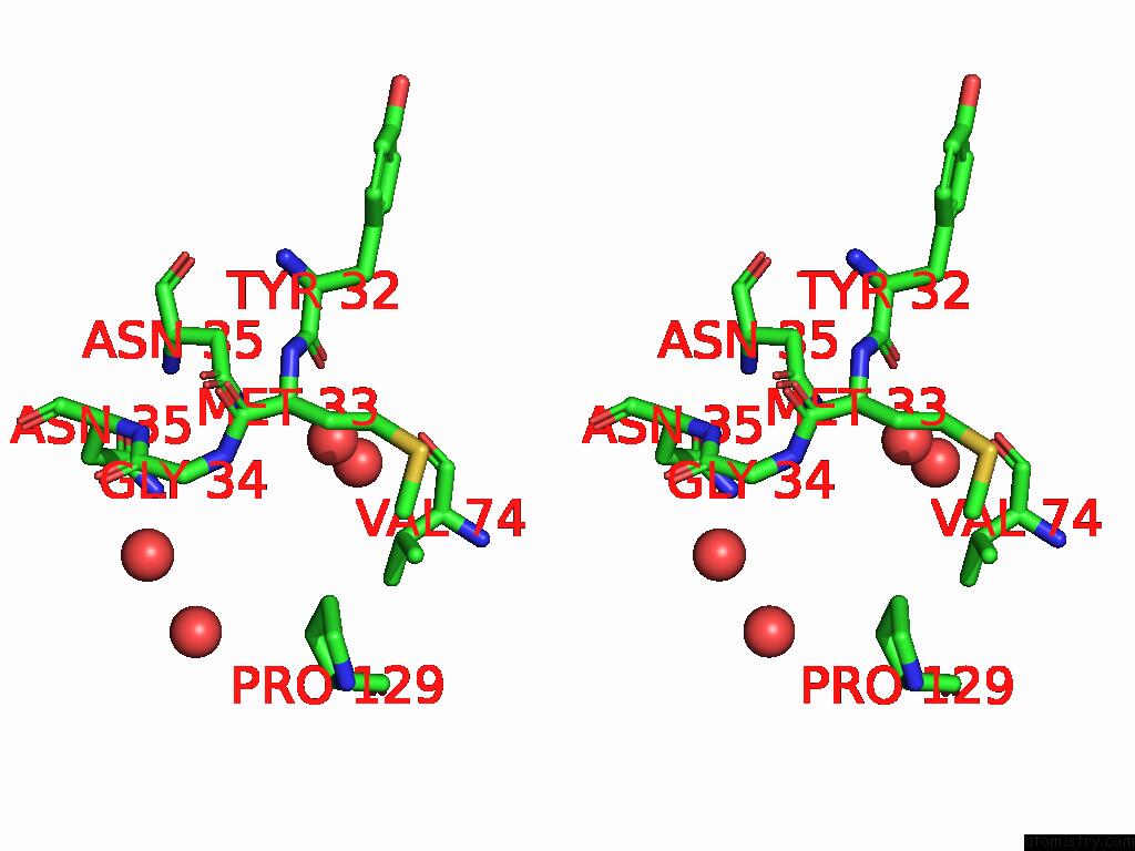
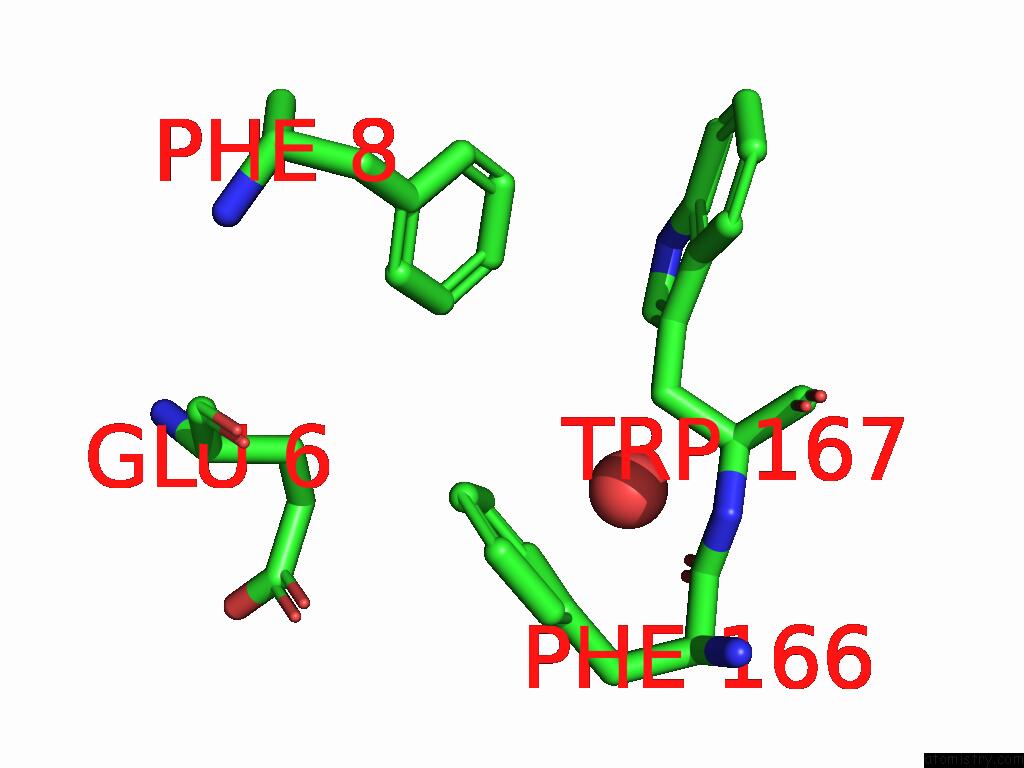
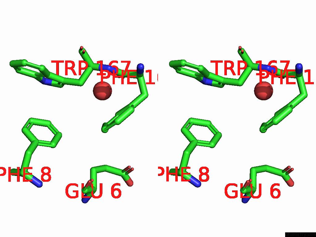
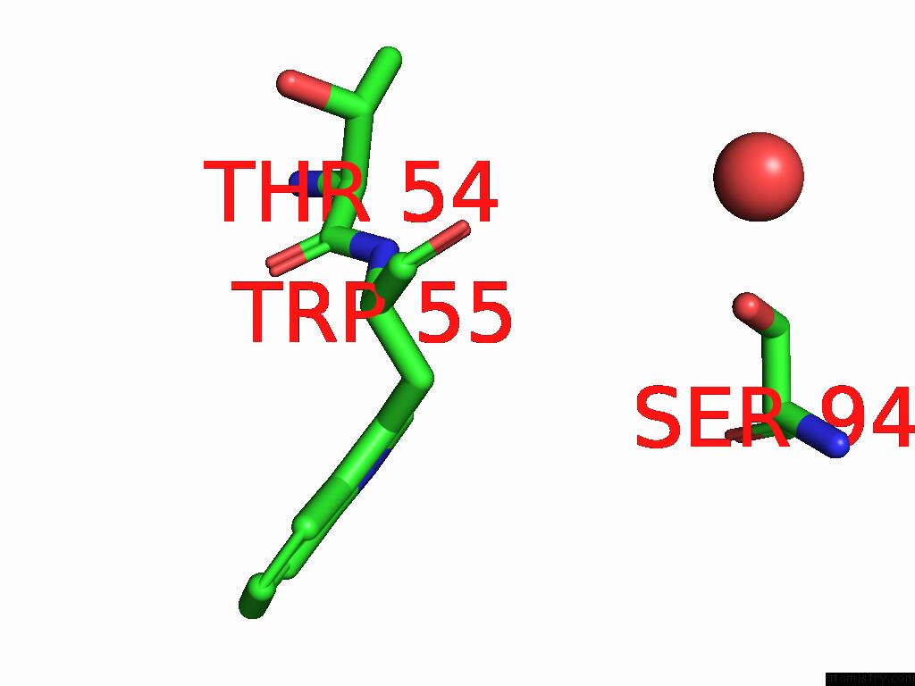
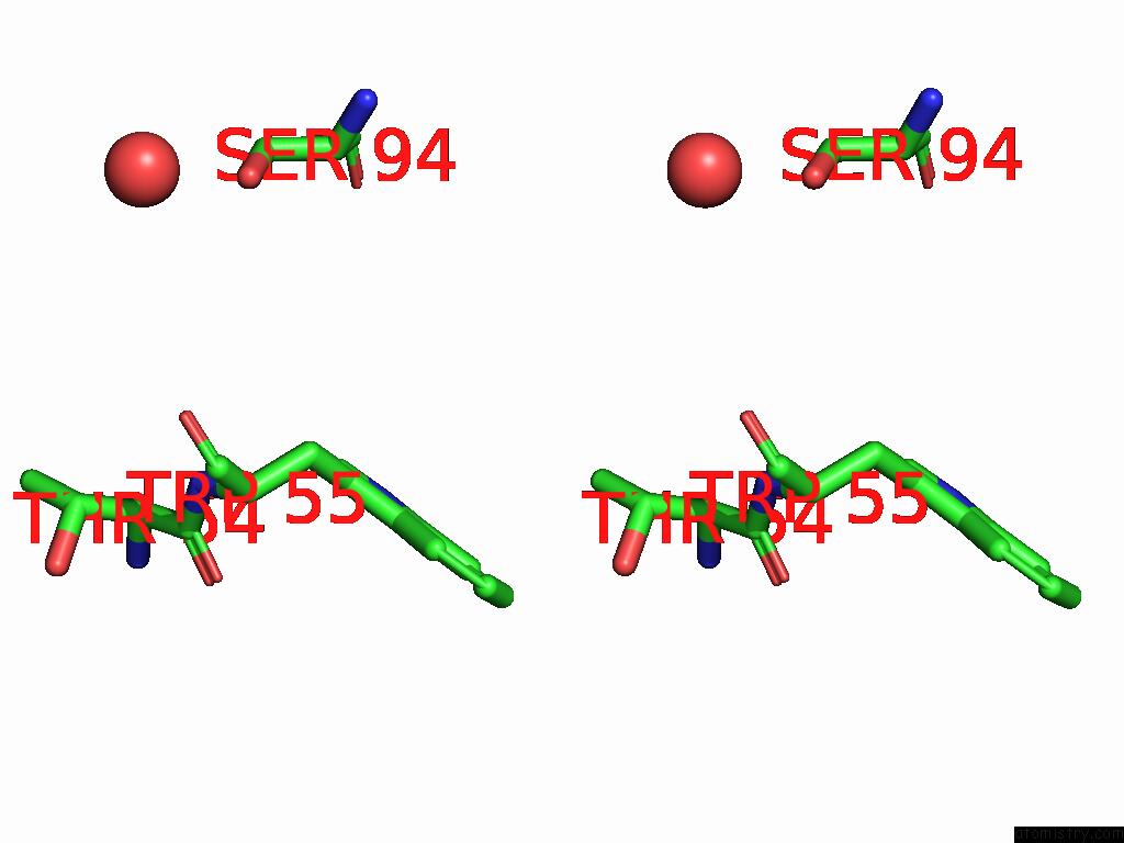
Iodine binding site 1 out of 7 in 9dq1
Go back to
Iodine binding site 1 out
of 7 in the Crystal Structure of Hrmj From Streptomyces Sp. Cfmr 7 (Hrmj-Ssc) Complexed with Manganese (II), 2-Oxoglutarate and 6-Nitronorleucine
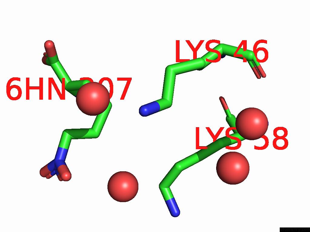
Mono view
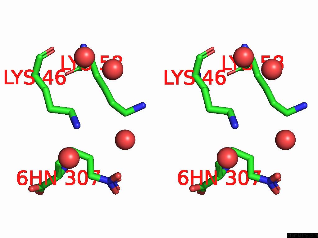
Stereo pair view
Mono view
Stereo pair view
A full contact list of Iodine with other atoms in the I binding
site number 1 of Crystal Structure of Hrmj From Streptomyces Sp. Cfmr 7 (Hrmj-Ssc) Complexed with Manganese (II), 2-Oxoglutarate and 6-Nitronorleucine within 5.0Å range:
|
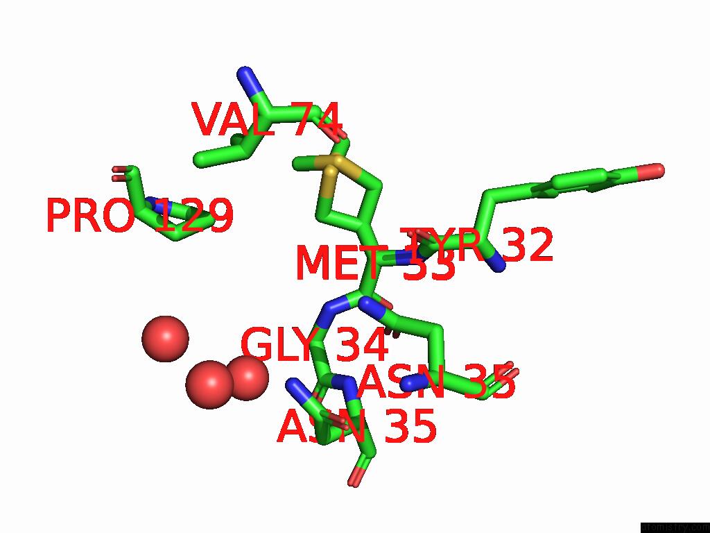
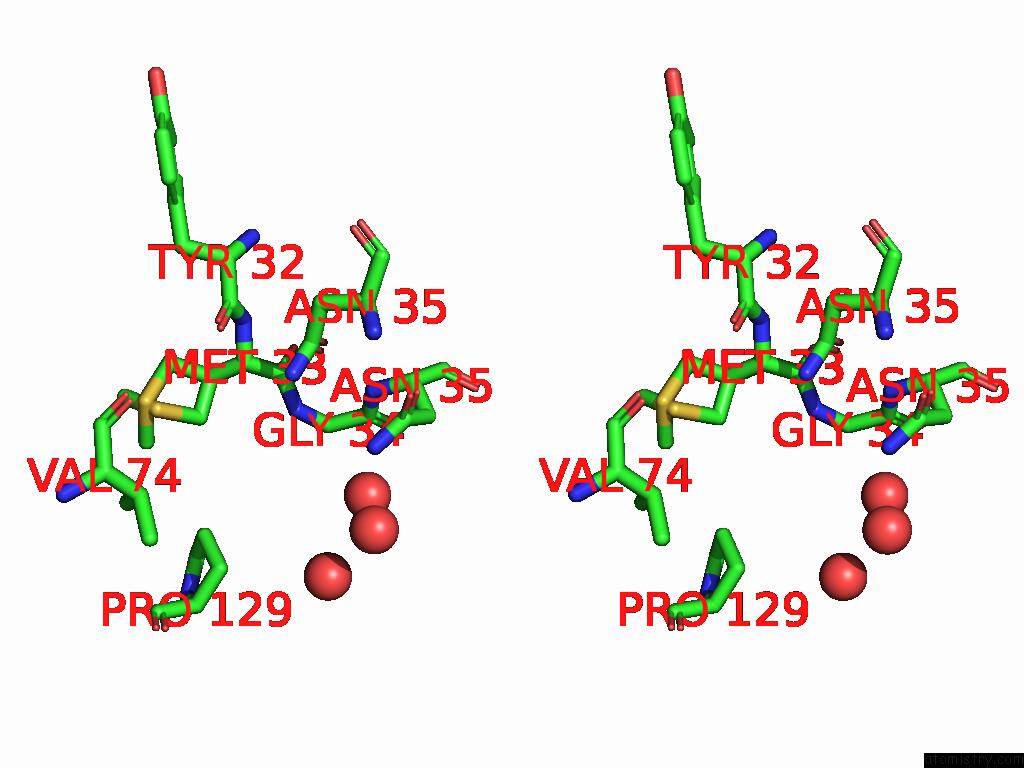
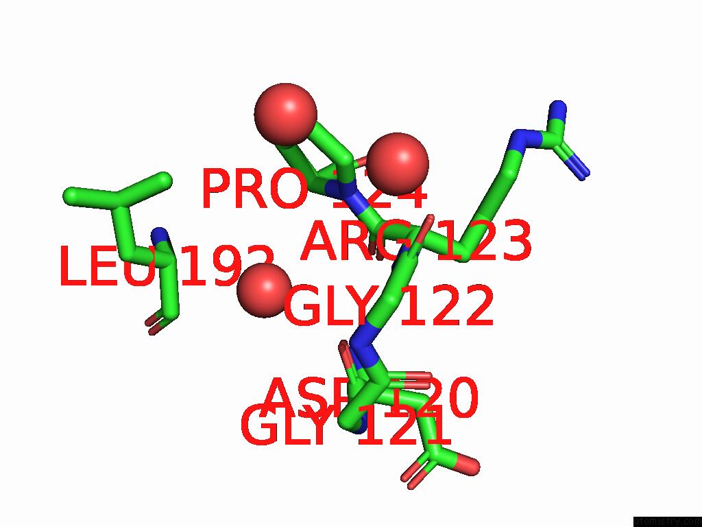
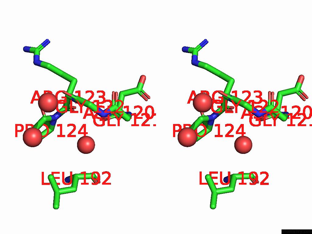
Iodine binding site 2 out of 7 in 9dq1
Go back to
Iodine binding site 2 out
of 7 in the Crystal Structure of Hrmj From Streptomyces Sp. Cfmr 7 (Hrmj-Ssc) Complexed with Manganese (II), 2-Oxoglutarate and 6-Nitronorleucine
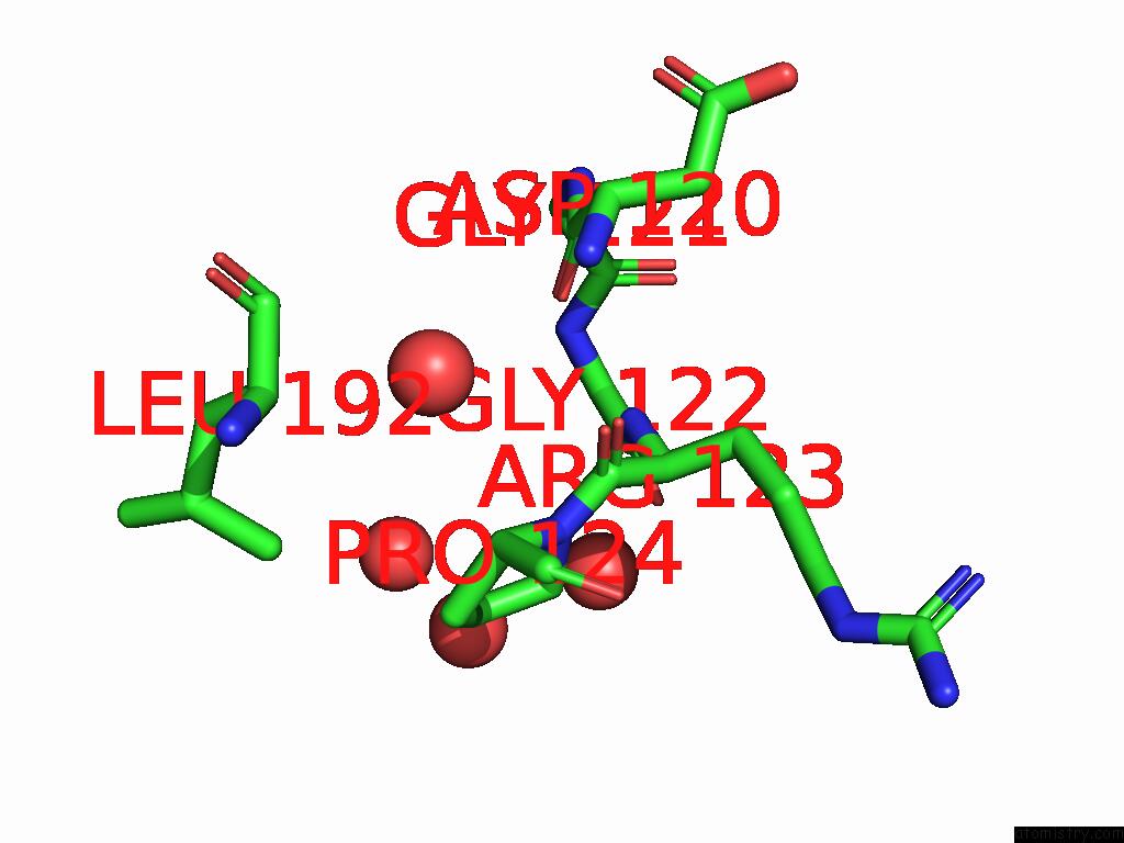
Mono view
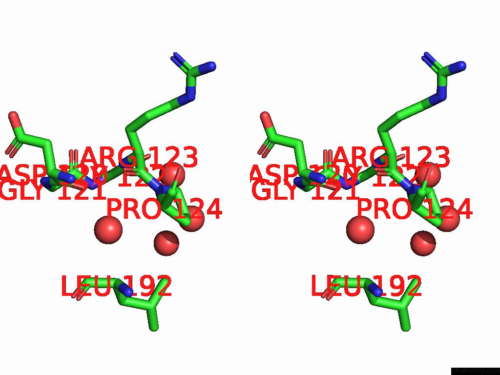
Stereo pair view
Mono view
Stereo pair view
A full contact list of Iodine with other atoms in the I binding
site number 2 of Crystal Structure of Hrmj From Streptomyces Sp. Cfmr 7 (Hrmj-Ssc) Complexed with Manganese (II), 2-Oxoglutarate and 6-Nitronorleucine within 5.0Å range:
|
Iodine binding site 3 out of 7 in 9dq1
Go back to
Iodine binding site 3 out
of 7 in the Crystal Structure of Hrmj From Streptomyces Sp. Cfmr 7 (Hrmj-Ssc) Complexed with Manganese (II), 2-Oxoglutarate and 6-Nitronorleucine
Mono view
Stereo pair view
Mono view
Stereo pair view
A full contact list of Iodine with other atoms in the I binding
site number 3 of Crystal Structure of Hrmj From Streptomyces Sp. Cfmr 7 (Hrmj-Ssc) Complexed with Manganese (II), 2-Oxoglutarate and 6-Nitronorleucine within 5.0Å range:
|
Iodine binding site 4 out of 7 in 9dq1
Go back to
Iodine binding site 4 out
of 7 in the Crystal Structure of Hrmj From Streptomyces Sp. Cfmr 7 (Hrmj-Ssc) Complexed with Manganese (II), 2-Oxoglutarate and 6-Nitronorleucine
Mono view
Stereo pair view
Mono view
Stereo pair view
A full contact list of Iodine with other atoms in the I binding
site number 4 of Crystal Structure of Hrmj From Streptomyces Sp. Cfmr 7 (Hrmj-Ssc) Complexed with Manganese (II), 2-Oxoglutarate and 6-Nitronorleucine within 5.0Å range:
|
Iodine binding site 5 out of 7 in 9dq1
Go back to
Iodine binding site 5 out
of 7 in the Crystal Structure of Hrmj From Streptomyces Sp. Cfmr 7 (Hrmj-Ssc) Complexed with Manganese (II), 2-Oxoglutarate and 6-Nitronorleucine
Mono view
Stereo pair view
Mono view
Stereo pair view
A full contact list of Iodine with other atoms in the I binding
site number 5 of Crystal Structure of Hrmj From Streptomyces Sp. Cfmr 7 (Hrmj-Ssc) Complexed with Manganese (II), 2-Oxoglutarate and 6-Nitronorleucine within 5.0Å range:
|
Iodine binding site 6 out of 7 in 9dq1
Go back to
Iodine binding site 6 out
of 7 in the Crystal Structure of Hrmj From Streptomyces Sp. Cfmr 7 (Hrmj-Ssc) Complexed with Manganese (II), 2-Oxoglutarate and 6-Nitronorleucine
Mono view
Stereo pair view
Mono view
Stereo pair view
A full contact list of Iodine with other atoms in the I binding
site number 6 of Crystal Structure of Hrmj From Streptomyces Sp. Cfmr 7 (Hrmj-Ssc) Complexed with Manganese (II), 2-Oxoglutarate and 6-Nitronorleucine within 5.0Å range:
|
Iodine binding site 7 out of 7 in 9dq1
Go back to
Iodine binding site 7 out
of 7 in the Crystal Structure of Hrmj From Streptomyces Sp. Cfmr 7 (Hrmj-Ssc) Complexed with Manganese (II), 2-Oxoglutarate and 6-Nitronorleucine
Mono view
Stereo pair view
Mono view
Stereo pair view
A full contact list of Iodine with other atoms in the I binding
site number 7 of Crystal Structure of Hrmj From Streptomyces Sp. Cfmr 7 (Hrmj-Ssc) Complexed with Manganese (II), 2-Oxoglutarate and 6-Nitronorleucine within 5.0Å range:
|
Reference:
Y.C.Zheng,
X.Li,
L.Cha,
J.C.Paris,
C.Michael,
R.Ushimaru,
Y.Ogasawara,
I.Abe,
Y.Guo,
W.C.Chang.
Comparison of A Nonheme Iron Cyclopropanase with A Homologous Hydroxylase Reveals Mechanistic Features Associated with Distinct Reaction Outcomes. J.Am.Chem.Soc. 2025.
ISSN: ESSN 1520-5126
PubMed: 39901767
DOI: 10.1021/JACS.4C17741
Page generated: Sat Aug 9 00:41:00 2025
ISSN: ESSN 1520-5126
PubMed: 39901767
DOI: 10.1021/JACS.4C17741
Last articles
K in 3B0GK in 3B0J
K in 3AY9
K in 3AHS
K in 3AOP
K in 3ATV
K in 3AFJ
K in 3ACT
K in 3A3Y
K in 3A45