Iodine »
PDB 1gjd-1lij »
1hc9 »
Iodine in PDB 1hc9: Alpha-Bungarotoxin Complexed with High Affinity Peptide
Protein crystallography data
The structure of Alpha-Bungarotoxin Complexed with High Affinity Peptide, PDB code: 1hc9
was solved by
M.Harel,
R.Kasher,
J.L.Sussman,
with X-Ray Crystallography technique. A brief refinement statistics is given in the table below:
| Resolution Low / High (Å) | 23.47 / 1.80 |
| Space group | C 2 2 21 |
| Cell size a, b, c (Å), α, β, γ (°) | 42.042, 153.356, 73.263, 90.00, 90.00, 90.00 |
| R / Rfree (%) | 20.2 / 23.5 |
Iodine Binding Sites:
The binding sites of Iodine atom in the Alpha-Bungarotoxin Complexed with High Affinity Peptide
(pdb code 1hc9). This binding sites where shown within
5.0 Angstroms radius around Iodine atom.
In total 2 binding sites of Iodine where determined in the Alpha-Bungarotoxin Complexed with High Affinity Peptide, PDB code: 1hc9:
Jump to Iodine binding site number: 1; 2;
In total 2 binding sites of Iodine where determined in the Alpha-Bungarotoxin Complexed with High Affinity Peptide, PDB code: 1hc9:
Jump to Iodine binding site number: 1; 2;
Iodine binding site 1 out of 2 in 1hc9
Go back to
Iodine binding site 1 out
of 2 in the Alpha-Bungarotoxin Complexed with High Affinity Peptide
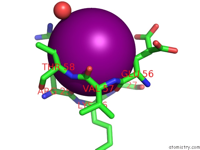
Mono view
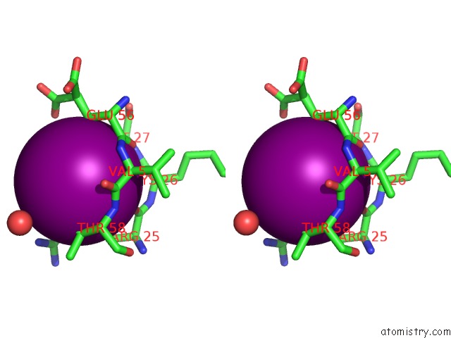
Stereo pair view
Mono view
Stereo pair view
A full contact list of Iodine with other atoms in the I binding
site number 1 of Alpha-Bungarotoxin Complexed with High Affinity Peptide within 5.0Å range:
|
Iodine binding site 2 out of 2 in 1hc9
Go back to
Iodine binding site 2 out
of 2 in the Alpha-Bungarotoxin Complexed with High Affinity Peptide
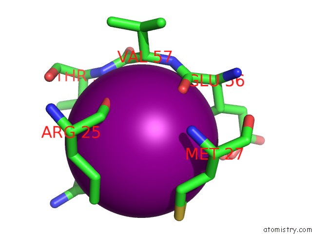
Mono view
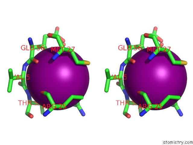
Stereo pair view
Mono view
Stereo pair view
A full contact list of Iodine with other atoms in the I binding
site number 2 of Alpha-Bungarotoxin Complexed with High Affinity Peptide within 5.0Å range:
|
Reference:
M.Harel,
R.Kasher,
A.Nicolas,
J.M.Guss,
M.Balass,
M.Fridkin,
A.B.Smit,
K.Brejc,
T.K.Sixma,
E.Katchalski-Katzir,
J.L.Sussman,
S.Fuchs.
The Binding Site of Acetylcholine Receptor As Visualized in the X-Ray Structure of A Complex Between Alpha-Bungarotoxin and A Mimotope Peptide. Neuron V. 32 265 2001.
ISSN: ISSN 0896-6273
PubMed: 11683996
DOI: 10.1016/S0896-6273(01)00461-5
Page generated: Fri Aug 8 11:52:10 2025
ISSN: ISSN 0896-6273
PubMed: 11683996
DOI: 10.1016/S0896-6273(01)00461-5
Last articles
I in 4J3WI in 4J3V
I in 4IZG
I in 4J3T
I in 4J3S
I in 4J2V
I in 4J1O
I in 4J0R
I in 4IWP
I in 4IY9